![]()
![]()
![]()
Use LEFT and RIGHT arrow keys to navigate between flashcards;
Use UP and DOWN arrow keys to flip the card;
H to show hint;
A reads text to speech;
197 Cards in this Set
- Front
- Back
Front (Term) |
Back (Definition) |
|
|
What makes up the perineum? |
Anus External genitalia |
|
|
What are the primary functions of the pelvis? |
- Bear weight of the upper body when sitting and standing - Transfer weight from the axial skeleton to the lower limbs for standing and walking - Provide attachment for muscles of locomotion and posture |
|
Front (Term) |
Back (Definition) |
|
|
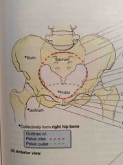
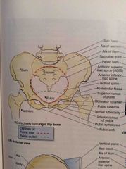
What bones make up the pelvic girdle? |
Left and Right hip bones Sacrum |
|
|
What makes up the hip bones? |
Ilium Ischium Pubic bone |
|
Front (Term) |
Back (Definition) |
|
|
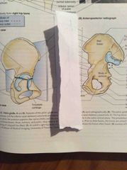
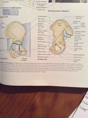
What are the boundaries of the pelvic inlet? |
Sacral ala promontories Arcuate line of the ilium Pectineal line along the superior ramus and body of the pubis |
|
|
What are the boundaries of the pelvic outlet? |
Pubic arch Ischial tuberosities Inferior margin of the sacrotuberous ligaments Tip of the coccyx |
|
|
What separates the greater and lesser pelvis? |
Pelvic inlet |
|
|
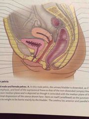
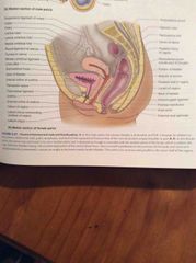
How is a female pelvis different to a male pelvis? |
Thinner and lighter Shallower and wider Wide, oval pelvic inlet Larger pelvic outlet Wide suprapubic angle Smaller acetabulum |
|
|
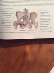
How is the sacrum attached to the ileal bones? |
Anterior sacroiliac ligaments Interosseus sacroiliac ligaments Posterior sacroiliac ligaments |
|
|
What is the sacrotuberous ligament? |
Passes from the posterior ilium and lateral sacrum/coccyx to the ischial tuberosity - this creates the sciatic foramen |
|
|
What is the sacrospinous ligament? |
Passes from lateral sacrum and coccyx to the ischial spine - subdivides the sciatic foramen into greater and lesser |
|
|
What kind of joint is the pubis symphysis? |
Secondary cartilagenous with an interpubic disc and surrounding superior and inferior pubic ligaments |
|
|
What direction are the facet joints of the lumbosacral joint? |
Facets on S1 face posteromedial. Facets on L5 face anterolateral. - prevents anterior slipping of the lumbar vertebra on the incline of the sacrum |
|
|
What reinforces the sacrococcygeal joint? |
Anterior and posterior sacrococcygeal ligaments |
|
|
Front (Term) |
Back (Definition) |
|
|
What are the “types” of pelvis? |
Android Gynaecoid Anthropoid Platypelloid |
|
|
What is the narrowest fixed distance through which a babies head must pass in the pelvis? |
AP diameter of the lesser pelvis (sacral promontory to pubic symphysis) |
|
|
What is the narrowest part of the pelvic canal? |
Interspinous distance (between the ischial spines) |
|
|
What injury is common in a fall from a height landing on ones feet? |
Fracture of femur through the acetabulum into the pelvic cavity |
|
|
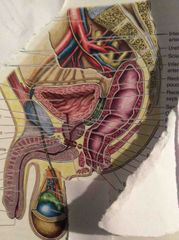
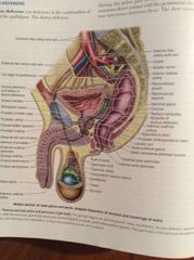
Pelvic cavity |
338 |
|
|
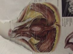
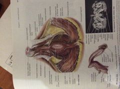
What makes up the walls of the pelvic cavity? |
- Anteroinferior = bodies and rami of the pubic bones - Lateral = right and left hip bones containing the obturator membrane and obturator internus muscle - Posterior = sacrum and coccyx and piriformis muscle - Pelvic floor = pelvic diaphragm |
|
|
What is the Pouch of Douglas? |
Rectouterine pouch - created by reflections of peritoneum covering the surface of the rectum and uterus |
|
|
What is the broad ligament of the uterus? |
A double-layered fold of peritoneum extending between the uterus and the lateral pelvic walls, which creates a partition between the lateral fossae of the bladder and rectum |
|
|
What is enclosed in the broad ligament? |
Uterine tubes Ovaries Ligaments of the ovaries Round ligaments |
|
|
What is the parietal pelvic fascia a continuation of? |
The transversalis and iliosoas fascias |
|
|
Front (Term) |
Back (Definition) |
|
|
Which muscles are most likely to tear during childbirth? |
Pubococcygeus Puborectalis |
|
|
Vadatur |
349 |
|
|
What makes up the pelvic diaphragm? |
Coccygeus muscle + fascia Levator any muscle + fascia |
|
|
Front (Term) |
Back (Definition) |
|
|
Front (Term) |
Back (Definition) |
|
|
What are the attachments of obturator internus? |
Pelvic surface of ilium and ischium to greater trochanter of femur |
|
|
What are the attachments of piriformis? |
Pelvic surface of S2-4 segments and superior margin of greater sciatic notch and sacrotuberous ligament to greater trochanter of femur |
|
|
What are the attachments of coccygeus muscle? |
Ischial spine to inferior end of sacrum and coccyx |
|
|
What are the muscles of levator ani? |
Pubococcygeus (Fibers to pro star, perineum and anus) Puborectalis Iliococcygeus |
|
|
Front (Term) |
Back (Definition) |
|
|
Which are the only intraperitoneal pelvic organs? |
Uterine tubes Ovaries |
|
|
What makes up the walls of the pelvic cavity? |
- Anteroinferior = bodies and rami of the pubic bones - Lateral = right and left hip bones containing the obturator membrane and obturator internus muscle - Posterior = sacrum and coccyx and piriformis muscle - Pelvic floor = pelvic diaphragm |
|
|
What is the parietal pelvic fascia a continuation of? |
The transversalis and iliosoas fascias |
|
|
Which muscles are most likely to tear during childbirth? |
Pubococcygeus Puborectalis |
|
|
Vadatur |
349 |
|
|
Front (Term) |
Back (Definition) |
|
|
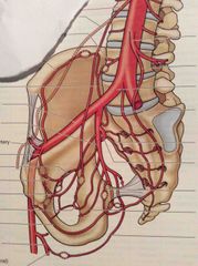
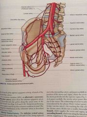
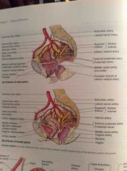
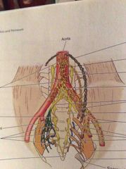
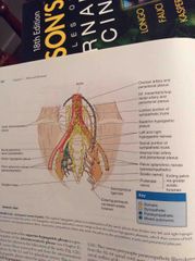
List the arteries supplying the pelvis. |
Gonadal Superior rectal (off IMA) Median sacral Anterior division of internal iliac; - umbilical - Superior and inferior vesical - obturator - Artery to ductus deferens and prostatic (men) - Uterine and vaginal (women) - internal pudendal - middle rectal - Inferior gluteal Posterior division of internal iliac - iliolumbar - Lateral sacral - Superior gluteal |
|
|
What are the 6 main arteries that enter the pelvis? |
2x internal iliac 2x ovarian (women) Median sacral Superior rectal |
|
|
Where does the common iliac artery bifurcate? |
Level between L5 and S1 |
|
|
Where does the internal iliac divide? |
At the superior edge of the greater sciatic foramen - divides into anterior and Posterior divisions |
|
|
What are the main branches of the anterior internal iliac artery? |
Umbilical Obturator - ilial and pubic branches Inferior vesicle (males) Uterine - vaginal, ovarian and tubal branches Middle rectal Internal pudendal - through the pudendal canal in the lateral wall of the ischioanal fossa Inferior gluteal |
|
|
What are the branches of the internal pudendal artery? |
Perineal artery Diesel artery of the penis/clitoris |
|
|
What makes up the pelvic diaphragm? |
Coccygeus muscle + fascia Levator any muscle + fascia |
|
|
What are the branches of the posterior division of the internal iliac? |
Iliolumbar - iliac and lumbar branches Lateral sacral - Superior and inferior Superior gluteal |
|
Front (Term) |
Back (Definition) |
|
|
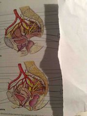
What is the course of the ovarian artery? |
Arises from the abdominal aorta, inferior to the renal artery but above IMA - Adheres to the parietal peritoneum and passes inferiorly on the posterior abdominal wall - enters the lesser pelvis, crossing the origin of the external iliac vessels - Runs medially and gives off an ovarian and tubal branch |
|
|
What is the superior rectal artery? |
The direct continuation of the IMA, which crosses the Left common iliac vessels and descends in the sigmoid mesocolon to the lesser pelvis |
|
|
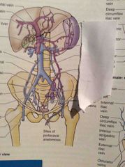
What level do the common iliac veins combine? |
L4/5 |
|
|
What are the 4 primary groups of LNs in the pelvis? |
1. External iliac - above the pelvic brim along the external iliac vessels 2. Internal iliac- around the anterior and posterior divisions of the internal iliac 3. Sacral - drain either to internal or common iliac nodes 4. Common iliac - Superior to the pelvis |
|
|
Front (Term) |
Back (Definition) |
|
|
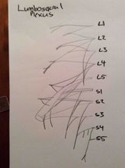
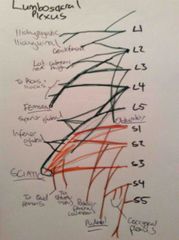
What are the 2 main nerve plexuses of the pelvis? |
Sacral Coccygeal - sit on the piriformis and coccygeus muscles |
|
|
What is the course of the obturator nerve? |
Arises from anterior rami of L2-4 of the lumbar plexus; - enters the lesser pelvis and runs in the extraperitoneal fat along the lateral wall of the pelvis to the obturator canal - goes through the obturator canal and divides into anterior and posterior parts - supplies the medial thigh muscles |
|
Front (Term) |
Back (Definition) |
|
|
What are the attachments of obturator internus? |
Pelvic surface of ilium and ischium to greater trochanter of femur |
|
|
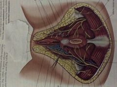
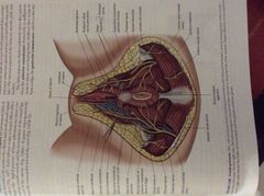
What is the course of the pudendal nerve? |
Leaves the sacral plexus via the greater sciatic foramen between piriformis and coccygeus - Hooks around the ischial spine and sacrospinous ligament - Enters the perineum through the lesser sciatic foramen Supplies the perineum and external genitalia |
|
|
What is the coccygeal plexus? |
Small network of nerve fibres formed by anterior rami of S4/5 and the coccygeal nerves - supplies coccygeus, levator ani and the sacrococcygeal joint |
|
|
Front (Term) |
Back (Definition) |
|
Front (Term) |

Back (Definition) |
|
|
What are the 4 routes for autonomic nerve entry into the pelvis? |
1. Sacral sympathetic trunks 2. Periarterial plexuses 3. Hypogastric plexus 4. Pelvis splanchnic nerves |
|
|
W |
Back (Definition) |
|
|
What is the pelvic pain line, and what does it represent?where |
Corresponds to the inferior limit of the peritoneum - Viscera Superior to the pain line (in contact with peritoneum) send pain afferent a via sympathetic channels - Viscera Inferior to the pain line send pain afferents via parasympathetic channels |
|
|
Where does the uterine artery cross the ureter? |
2 cm superior to the ischial spine |
|
|
What is the course of the ureters in the pelvis? |
Cross the bifurcation of the common iliac artery and pass over the pelvic brim - Run on the Lateral walls of the pelvis between the parietal peritoneum and the internal iliac arteries - Opposite the ischial spine, they curve anteromedially, Superior to levator ani and enter the bladder |
|
Front (Term) |
Back (Definition) |
|
|
What are the attachments of piriformis? |
Pelvic surface of S2-4 segments and superior margin of greater sciatic notch and sacrotuberous ligament to greater trochanter of femur |
|
|
Where is ureteric pain referred to? |
Ipsilateral lower quadrant, especially the groin |
|
W |
Back (Definition) |
|
|
What anchors the neck of the bladder? |
Lateral ligaments of the bladder Tendinous arch of the pelvic fascia |
|
|
Front (Term) |
Back (Definition) |
|
|
Front (Term) |
Back (Definition) |
|
|
How are the muscle fibers in the neck of the bladder different in males and females? |
Males - detrusor muscle contracts at the neck to form an internal urethral sphincter that tightens during ejaculation. Muscle fibers are continuous with the fibromuscular tissue of the prostate.
Female - muscle fibers in the neck are continuous with muscle fibers in the wall of the urethra |
|
|
What is the arterial supply to the bladder? What wh |
Branches of the internal iliac. - Superior vesical arteries - Males = Inferior vesical arteries - Females = Vaginal arteries |
|
|
What is the nerve supply to the bladder? |
Sympathetic = from inferior thoracic and upper lumbar SC levels to the pelvic plexus via hypogastric plexus
Parasympathetic = from sacral SC levels via pelvic splanchnic nerves and the inferior hypogastric plexus
Sensory = mostly follow parasympathetic, by superior bladder via sympathetic |
|
|
What are the 4 parts of the male urethra? |
- Intramural: through the neck of the bladder, surrounded by IUS - Prostatic: through anterior prostate, widest and most delicate part with a urethral crest and seminal colliculus (opening of ejaculatory ducts) - Membranous: through deep perineal pouch, surrounded by external sphincter and penetrates the perineal membrane - Spongy: through corpus spongiosum, receives input from bulbourethral glands |
|
Front (Term) |
Back (Definition) |
|
Front (Term) |
Back (Definition) |
|
|
What are the features of the female urethra? |
- 4cm long from internal urethral orifice to the external urethral orifice in the vestibule - Lies directly anterior to the vagina - Blood supply via internal pudendal and vaginal arteries - Nerve supply via vesical plexus and the pudendal nerve |
|
|
What are the flexures of the rectum? |
Sacral flexure (as it follows the curvature of the sacrum) Anorectal flexure (as it perforates the pelvic diaphragm through levator ani) |
|
|
What are the 3 lateral flexures of the rectum? |
Left = superior and inferior Right = intermediate |
|
|
What part of the rectum is directly superior to levator ani and anococcygeal ligament? |
Ampulla |
|
|
Which parts of the rectum are covered in peritoneum? |
Superior 1/3 = anterior and lateral parts covered Middle 1/3 = anterior part covered Inferior 1/3 = not covered by peritoneum |
|
|
What sits posterior to the rectum? |
S3-5 vertebrae Coccyx Anococcygeal ligament Median sacral vessels Inferior ends of the sympathetic trunks and sacral plexuses |
|
|
What is the arterial supply of the rectum? |
Superior rectal (off IMA) Middle rectal (off internal iliac) Inferior rectal ( off internal pudendal) |
|
|
What are the 2 parts of the rectal venous plexus? |
Internal - just deep to the mucosa of the anorectal junction External - external to the muscular wall of the rectum |
|
|
What is the nerve supply to the rectum? |
Sympathetic - lumbar SC via lumbar splanchnic nerves and hypogastric plexus
Parasympathetic - S2-4 via pelvic splanchnic and inferior hypogastric
Visceral - follow parasympathetic fibres |
|
|
What is a cystocoele? |
Collapse of the bladder into the anterior vaginal wall |
|
|
What are the muscles of levator ani? |
Pubococcygeus (Fibers to pro star, perineum and anus) Puborectalis Iliococcygeus |
|
|
Male g |
A |
|
|
Which are the only intraperitoneal pelvic organs? |
Uterine tubes Ovaries |
|
|
What is the Pouch of Douglas? |
Rectouterine pouch - created by reflections of peritoneum covering the surface of the rectum and uterus |
|
|
What is the broad ligament of the uterus? |
A double-layered fold of peritoneum extending between the uterus and the lateral pelvic walls, which creates a partition between the lateral fossae of the bladder and rectum |
|
What is enclosed in the broad ligament?
|
Uterine tubes
Ovaries Ligaments of the ovaries Round ligaments |
|
|
What are the features of the ductus deferens? |
- Continuation of the duct of the epididymis - Thick, muscular wall - Primary component of the spermatic cord - Joins the duct of the seminal gland to form the ejaculatory duct |
|
|
Where are the bulbourethral glands? |
Posterolateral to the intermediate part of the urethra, largely embedded within the external urethral sphincter |
|
|
What happens at orgasm in the male genitourinary system? |
Sympathetic action; - Contraction of the internal urethral sphincter - Stimulation of rapid peristalsis-like contractions of the ductus deferens - Contraction of and secretion from the seminal glands and prostate |
|
|
What is a vasectomy? |
Excision through the ductus deferens in the Superior part of the scrotum |
|
|
Which part of the prostate is spared in prostate surgery and why? |
The capsule - it contains much of the venous and nerve structures that enable sexual function and urinary control |
|
|
What is the vascular supply to the ovary? |
Ovarian artery through the suspensory ligament of the ovary, which becomes continuous with he mesovarium. |
|
|
What are the 4 parts of the uterine tube? |
Fimbria and infundibulum Ampulla (site of fertilization) Isthmus Uterine part |
|
|
What is the arterial supply of the uterine tube? |
Branch from the ovarian artery Ascending branch of the uterine artery |
|
|
Front (Term) |
Back (Definition) |
|
|
What is the usual position of the uterus? |
Anteverted and antiflexed - lies over the bladder |
|
|
What are the layers of the uterine wall? |
Perimetrium - serosa Myometrium - smooth muscle Endometrium - inner mucus coat |
|
|
What is the course of the ductus deferens? |
Begins in the tail of the epididymis - ascends posterior to the testis - penetrates the anterior abdominal wall via the inguinal canal - crosses over the external iliac vessels and enters the pelvis - passes along the lateral wall of the pelvis, external to the peritoneum - crosses superior to the ureter to reach the Fundus of the bladder |
|
|
What are the remnants of the ovarian gubernaculum? |
Ligament of the ovary Round ligament of the uterus |
|
|
What are the parts of the broad ligament? |
Mesovarium Mesosalpinx Mesometrium Suspension ligament |
|
|
What are the supports that anchor the cervix? |
Cardinal ligaments - from supravaginal cervix and lateral fornices to the lateral walls of the pelvis
Uterosacral ligaments - pass superiorly and slightly posteriorly from the sides of the cervix to the middle of the sacrum |
|
|
What is the vagina? |
A distensible musculomembranous tube that extends from the middle cervix to the vaginal orifice in the vestibule |
|
|
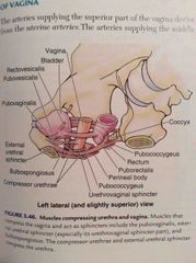
What 4 muscles compress the vagina and act as sphincters? |
Pubovaginalis External urethral sphincter Urethrovaginal sphincter Bulbospongiosus |
|
|
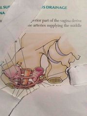
What is the arterial supply of the vagina? |
Superior - from uterine Middle and inferior - from vaginal and internal pudendal |
|
|
What is the somatic innervation of the vagina? |
Inferior 1/5 - Deep perineal nerve (branch of internal pudendal) |
|
|
What is the nerve supply of the superior vagina? |
Uterovaginal nerve plexus - from the inferior hypogastric plexus |
|
|
What is a hysterosalpingogram? |
Visualization of tubal patency by injection of a radio-opaque water soluble material into the uterus and tubes via the external os and observation under radiography |
|
|
What is the seminal gland? |
An elongated structure that lies between the Fundus of the bladder and the rectum. Secretes a thick alkaline fluid with fructose and a coagulating agent. |
|
|
What is the cause of a bicornate uterus? |
Incomplete fusion of the embryonic paramesonephric ducts |
|
|
What is Hegar sign? |
Softening of the uterine isthmus causing the cervix to feel separated from the body of the uterus - early sign of pregnancy |
|
|
What can the vagina form fistulas with? |
Bladder Urethra Rectum Perineum |
|
|
What is the artery supply of the seminal glands? |
Arteries derived from the inferior vesical and middle rectal arteries |
|
|
Where do the ejaculatory ducts empty into? |
The posterior prostatic urethra, through slit-like openings in the utricle of the seminal colliculi |
|
|
What is the composition of the prostate? |
Primarily glandular Surrounded by a fibromuscular capsule |
|
|
What are the lobes of. the prostate? |
- Isthmus: anterior lobe that is fibromuscular representing a continuation of the external urethral sphincter - Right and left lobes: separated anteriorly by the isthmus and posteriorly by a longitudinal furrow |
|
|
Where do the prostatic ducts open? |
Into prostatic sinuses, which is on either side of the seminal colliculi on the posterior wall of the prostatic urethra |
|
|
What is the arterial supply of the prostate? |
Branches of the inferior vesical, internal pudendal and middle rectal arteries |
|
|
What is the lymph drainage of the ureters? |
Superior part - to external iliac LNs Inferior part - to internal iliac LNs |
|
|
What is the lymph drainage of the bladder? |
Superolateral = external iliac LNs Fundus and neck = internal iliac LNs |
|
|
What is the lymph drainage of the rectum? |
Superior half = via inferior mesenteric LNs to pararectal region Inferior half = direct into the sacral LNs or internal iliac LNs |
|
|
What LNs does the external genitalia drain to? |
Inguinal LNs |
|
|
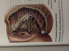
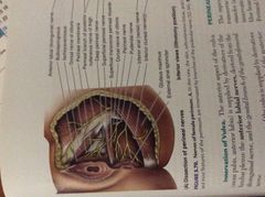
What are the boundaries of the perineum? |
Pubic symphysis annteriorly Ischiopubic rami + ischial rami anterolaterally Ischia tuberosities laterally Sacrotuberous ligament posterolaterally Inferiormost sacrum and coccyx posteriorly |
|
|
How is the perineum divided? |
Anterior (urogenital) triangle Posterior (anal) triangle |
|
|
What is at the midpoint of the perineum? |
The perineal body - an irregular mass containing collagenous and elastic fibres and skeletal and smooth muscle |
|
|
What muscle fibres converge in the perineal body? |
Bulbospongiosus External anal sphincter Superficial and deep transverse perineal muscles Slips of muscle from EAS, levator ani, rectum |
|
|
What is the urogenital comprised of? |
The perineal membrane - a thin sheet of tough fascia, which is perforated by the urethra and vagina |
|
|
|
|
|
What are the layers of fascia in the perinuem? |
Superficial fatty layer Deep membranous layer Deep perineal fascia |
|
|
What is the superficial perineal pouch?
|
A potential space between perineal fascia and the perineal membrane. contains; - Males: root of the penis, ischocavernosus, bulbospongiosus, spongy urethra, superficial transverse perineal mucscles, deep perineal branches if internal pudendal a/v/n - Females: clitoris, bulbs of the vestibule, greater vestibular glands, superficial transverse perineal muscles, perineal branches of internal pudendal a/n/v |
|
|
|
|
|
What is contained in the ischiorectal fossae? |
Fat bodies, which occupy space, but are readily displaced to permit descent and expansion of the anal canal during passage of faeces |
|
|
What is the pudendal canal? |
A horizontal passageway within the obturator fascia that covers the medial aspect of the obturator internus and lines the lateral wall of the ischiorectal fossa |
|
|
|
|
|
What is the function of the internal pudendal n/a/v? |
Supply to the perineum - distal rectal a/v - perineal n/a/v - dorsal arteryy of the penic/clitoris |
|
|
What are the branches of the perineal nerve? |
Superficial --> posterior scrotal/labial branches Deep --> supplies muscles of the deep and superficial perineal pouches, skin of the vestibule, inferior mucosa of the vagina |
|
|
Where does the anal canal begin? |
Where the ampulla of the rectum narrows at the level of the puborectalis (creates a sling) |
|
|
What are the 2 sphincters of the anal canal? |
Internal = involuntary, thickening of the circular muscular layer, tonically contracted External = large, voluntary sphincter thta forms a broad band on each side of the inferior 2/3 of the anal canal |
|
|
What nerves supply the external anal sphincter? |
S4 via inferior rectal nerve |
|
|
What is the pectinate line? |
Demarcation in the anal canal above and below which there is different arterial supply, innervation and lymph/venous drainage |
|
|
What is the internal structure of the anal canal? |
Superior half has longitudinal anal columns These terminate at the anorectal junction Inferior ends of the anal column are joined by anal valves which have small superior recesses (anal sinuses) |
|
|
What is included in the male urogenital triangle? |
External genitalia - penis, scrotum, distal urethra Perineal muscles |
|
|
What is the course of the membranous urethra? |
Begins at the apex of the prostate - traverses the deep perineal pouch, surrounded by the external urethral sphincter - Penetrates the perineal membrane - Ends as the urethra enters the bulb of the penis |
|
|
What is the course of the spongy urethra? |
Begins at the distal end of the the intermediate urethra, at the bulb of the penis - ends at the external urethral orifice |
|
|
Where is the lumen of the spongyg urethra expanded? |
- Bulb: forms the intrabulbar fossa - Glans: forms the navicular fossa |
|
|
Where do the bulbourethra glands open into? |
The proximal part of the spongy urethra |
|
|
|
|
|
What is the arterial, venous and lymph supply of the membranous and spongy urethra? |
Artery = branches of dorsal artery of the penis Vein = accompany artery Lymph; - Membranous to internal iliac LNs - Spongy to deep inguinal LNs |
|
|
What is the innervation of the distal male urethra? |
Membranous = via prostatic nerve plexus Spongy = dorsal nerve of the penis (branch of pudendal) |
|
|
What is the scrotal raphe continuous with? |
Anteriorly = penile raphe on the ventral surface Posteriorly = perineal raphe in the perineum |
|
|
What separates the 2 testes in the scrotum? |
A prolongation off the dartos fascia - septum of the scrotum |
|
|
What is the artery, nerve, lymph, vein supply to the scrotum? |
Artery = anterior scrotal arteries (off exterenal pudendal) and posterior scrotal arteries (off internal pudendal) Vein = accompany arteries to external pudendal vein Lymph = drain to the superficial inguinal LNs Nerve = anterior from the lumbar plexus (anterior scrotal nerves), posterior from the superficial perineal branches of the pudendal nerve (posterior scrotal nerves) |
|
|
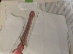
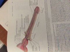
What are the parts of the penis? |
Root Body Glans |
|
|
What is the erectile tissue of the penis? |
2x corpora cavernosa dorsally 1x corpus spongiosum ventrally |
|
|
What are the CT layers of the penis? |
- Each cavernous body is covered by a tunica albuginea - Deep fascia of the penis (continuation of deep perineal fascia) covers the erectile tissue |
|
|
What do the copora cavernosa give rise to procimally? |
The crura of the penis |
|
|
What makes up the root of the penis? |
Crura Bulb Ischiocavernosus and bulbospongiosus muscles |
|
|
Where does the root of the penis sit? |
Between the perineal membrane superiorly and the deep perineal fascia inferiorly |
|
|
What is the suspensory ligament of the penis? |
Condensation of deep fascia that arises from the anterior surface of the pubic symphysis - passes inferiorly and splits to form a sling that is attached to the deep fascia of the penis |
|
|
|
|
|
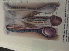
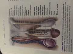
What is the arterial supply of the penis? |
Branches of the internal pudendal arteries; - Dorsal arteries of the penis - run either side of the deep dorsal vein - Deep arteries of the penis - run in the cavernosa - Arteries of the bulb of the penis - posterior supply - Superficial and deep branches of pudendal supply the penile skin |
|
|
What is the venous and lymphatic drainage of the penis? |
Venous = deep dorsal vein of the penis drains into the prostatic venous plexus. Skin drains via superficial external pudendal vein. Lymph; - Skin to superficial inguinal LNs - Proximal urethra to internal iliac LNs - Distal urethra to deep inguinal LNs |
|
|
What is the innervation of the penis? |
S2-4 Dorsal nerve of the penis - a terminal branch of the pudendal nerve |
|
|
|
|
|
What are the superficial perineal muscles? |
Superficial transverse perineal muscle Bulbospongiosus Ischiocavernous |
|
|
|
|
|
Which part of the urethra is most likely to be damaged during catheterisation? |
Thin segment of membranous urethra outside the bulb |
|
|
What is the narrowest and least distensible part of the male urethra? |
External urethral orifice |
|
|
Which testis is normally lower? |
Left |
|
|
What is a hypospadia? |
Congenital anomaly of the penis in which the external urethral orifice is on the ventral aspect of the glans penis |
|
|
What is the female external genitalia? |
Mons pubis Labia majora (enclosing pudendeal cleft) Labia minora (enclosing vestibule) Clitoris Bulb of vestibule Greater and lesser vestibular glands |
|
|
|
|
|
What are the 2 "ends" of the labia majora? |
Anterior commissure Posterior commissure (fuses with perineal body) |
|
|
What opens into the vestibule of the vagina? |
External urethral orifice Vagina |
|
|
What are the 2 "'ends" of the labia minora? |
Anterior = frenulum and prepuce of the clitoris Posterior = frenulum (foruchette) in virgins |
|
|
What is the clitoris composed of? |
Root and body Body contains 2 crura, 2 corpora cavernosa and the glans of the clitoris |
|
|
What do the crura of the clitoris attach to? |
The inferior pubic rami and perineal membrane |
|
|
What are the bulbs of the vestibule? |
Paired masses of elongated erectile tissue that lie along the sides of the vaginal orifice, deep to the labia minora and immediately inferior to the perineal membrane |
|
|
What are the vestibular glands? |
Bartholin glands - Mucus-secreting glands that lie on each side of the vestibule in the superficial perineal pouch |
|
|
What is the artery, vein and nerve supply to the vulva? |
Artery = external and internal pudendal arteries Veins = drain to the internal pudendal vein Nerve; - Anterior = anterior labial nerves from te ilioinguinal nerve and genital branch of genitofemoral - Posterior = perineal branch of the posterior cutaneous nerve of the thigh and pudendal nerve |
|
|
|
|
|
What are the superficial perineal muscles of the female? |
Superficial transverse perineal muscle Ischiocavernous Bulbospongiosus |
|
|
Where is a pudendal nerve block performed? |
Where the pudendal nerve crosses the lateral aspect of the sacrospinous ligament, near its attachment to the ischial spine |