![]()
![]()
![]()
Use LEFT and RIGHT arrow keys to navigate between flashcards;
Use UP and DOWN arrow keys to flip the card;
H to show hint;
A reads text to speech;
50 Cards in this Set
- Front
- Back
|
Short lateral rotator of the thigh |
Pifimoris |
|
|
Short lateral rotator of the thigh |
Obterutor internus |
|
|
Short lateral rotator of the thigh |
Inferior and superior gemelae |
|
|
Gluteal maximus origin |
Sacrum Behind posterior iliac line Sacrutuberus ligament |
|
|
Gluteal maximus insertion |
Superfecialy 2/3 iliotibial tract Deeply 1/3 gluteal line |
|
|
Inferior Extensor Retinaculum Insertion |
Upper .medial malleolus Lower .Medial cuneiform and Navicular |
|
|
Flexor Retinaculum |
Extends from medial malleolus to calcaneus |
|
|
Inferior Fibular Retinaculum |
Lateral calcanus to 5 metatarsal |
|
|
Superior Fibular Retinaculum |
Lateral malleolus to calcaneus |
|
|
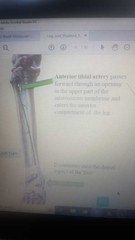
Popliteal artery site of branch |
Inferior border of Popliteus muscle |
|
|
Popliteal artery branches |
Anterior Tibial artery Posterior Tibial artery |
|
|
Anterior Tibial artery course in leg |
|
|
|
Posterior Tibial artery course |
|
|
|
Sciatic nerve branches |
Common Fibular nerve Tibial nerve |
|
|
Common Fibular nerve branches |
Deep Fibular nerve Superficial Fibular nerve |
|
|
Common Fibular nerve ending point |
Neck of Fibula |
|
|
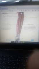
Muscles of Lateral compartment of leg supplies by |
Superficial Fibular nerve |
|
|
Muscles of Anterior compartment of the leg supplies by |
Deep Fibular nerve |
|
|
Muscles of Posterior compartment of leg supplies by |
Tibial nerve |
|
|
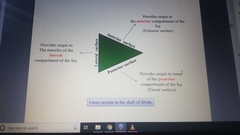
Role of Fibula in leg muscles |
|
|
|
Hallucis |
Big toe |
|
|
Digitorum |
Lateral 4 toes |
|
|
Peroneal |
Fibular |
|
|
Tibialis Anterior origin |
Shaft of tibia and interosseus memb |
|
|
Tibialis insertion |
Medial Cuniform and Base of 1 metatarsal bone |
|
|
Extensor Hallucis Longus origib |
Anterior surface of shaft of fibula |
|
|
Extensor Hallucis Longus insertion |
Base of Distal phalanx of big toe |
|
|
Extensor digitorum Longus origin |
Shaft of fibula and lateral condyl of tibia |
|
|
Extensor Digitorum Longus insertion |
Extensor expansion into bases of middle and distal phalanges of lateral four toes |
|
|
knee joint articulating surfaces |
between Femur and tibia between patella and patellar surface of femur |
|
|
knee joint tibiofemoral type |
modified synovial hinge |
|
|
patellofemoral joint type |
plane gliding |
|
|
capsule of knee joints |
|
|
|
ligaments of knee |
extracapsular;- 1. ligamentum patellae 2. lateral collateral ligament 3. medial collateral ligament 4. oblique popliteal ligament intracapsular 1.anterior cruciate ligament 2. posterior cruciate ligament |
|
|
the cruciate ligaments role |
the main bond bet the femur and tibia throughout the joint range of movement |
|
|
anterior cruciate ligament attachments |
anterior intercondylar area of the tibia and passes upward backward and laterally to be attached to the posterior part of the lateral wall of intercondylar fossa |
|
|
the anteror cruciate ligament role |
restricts the anterior displacement of tibia relative to the femur |
|
|
posterior cruciate ligament attachment |
attached to the posterior intercondylar area of the tibia and passes upward forward medially to be attached to anterior part of the medial wall of the intercondylar fossa |
|
|
posterior cruciate ligament role |
restricts posterior displacement of tibia relative to the femur |
|
|
anterior drawer sign |
forward (ACL) |
|
|
posterior drawer sign |
backward (PCL) |
|
|
lateral collateral ligament |
cord like between fibula and lateral epicondyle of femur it is Separated from lateral meniscus ligament |
|
|
medial collateral ligament |
flat band like bet above medial epicondyle of femur and below upper medial surface of tibia Attached firmly to the edge of medial meniscus |
|
|
popliteus tendon location |
intervenes between the capsule and lateral meniscus |
|
|
function of menisci |
1. deepen the articular surfaces of tibia condyles to receive the convex femoral condyles
2. cushion between two bones |
|
|
which is larger medial or lateral menisci |
medial |
|
|
medial menisci is relatevly immobile |
attached medially to the medial collateral ligament and capsule of the knee joint |
|
|
reason of the lateral condyle of femur is bit longer than the medial
|
helps preventing the lateral dislocation of patella |
|
|
bursae of knee |
communicate with joint cavity suprapatellar bursa popliteal bursa no communicate with joint cavity prepatellar bursa (housemaids knee) suparficial infrapatellar bursa deep infrapatellar bursa anserinus bursa subtendinous bursa of biceps femoris sartorius gastrocnemius iliotibial tract |
|
|
Inguinal ligament formed by |
External Abdominal Oblique Aponeurosis |