![]()
![]()
![]()
Use LEFT and RIGHT arrow keys to navigate between flashcards;
Use UP and DOWN arrow keys to flip the card;
H to show hint;
A reads text to speech;
15 Cards in this Set
- Front
- Back
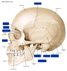
Skull: Lateral View
-Styloid process of the temporal bone -Mastoid process of the temporal bone -Squamous portion of the temporal bone -External acoustic meatus -Ethmoid bone -Supraorbital foramen -Infraorbital foramen -Sphenoid (greater wing) -Coronal suture |
-Styloid process of the temporal bone -Mastoid process of the temporal bone -Squamous portion of the temporal bone -External acoustic meatus -Ethmoid bone -Supraorbital foramen -Infraorbital foramen -Sphenoid (greater wing) -Coronal suture |
|
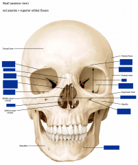
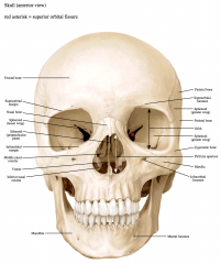
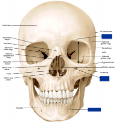
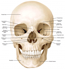
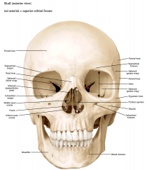
Skull: Anterior View
-Supraorbital margin -Infraorbital margin -Nasal bone -Mental Foramen -Ethmoid (perpendicular plate) -Supraorbital foramen -Orbit -Vomer -Sphenoid (Greater Wing) -Sphenoid (Lesser Wing) -Infraorbital Foramen -Piriform Aperture |
-Supraorbital margin -Infraorbital margin -Nasal bone -Mental Foramen -Ethmoid (perpendicular plate) -Supraorbital foramen -Orbit -Vomer -Sphenoid (Greater Wing) -Sphenoid (Lesser Wing) -Infraorbital Foramen -Piriform Aperture |
|
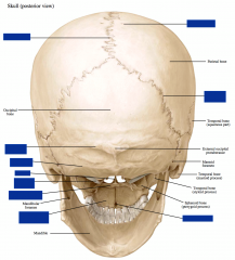
Skull: Posterior View
-Parietal foramen -Superior nuchal line -Inferior nuchal line -Vomer -Lamboid Suture -Incisive Foramen -Sagittal Suture -Occipital Condyle -Palantine bone (horizontal plate) -Palantine process of the Maxilla |
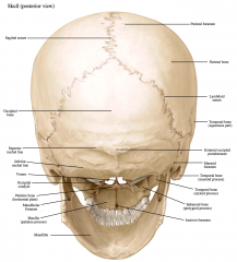
-Parietal foramen -Superior nuchal line -Inferior nuchal line -Vomer -Lamboid Suture -Incisive Foramen -Sagittal Suture -Occipital Condyle -Palantine bone (horizontal plate) -Palantine process of the Maxilla |
|
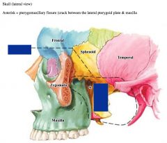
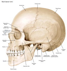
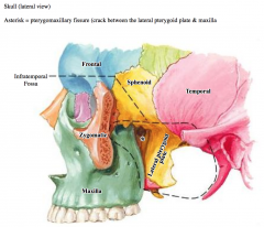
Skull: Lateral View 2
-Infratemporal fossa -Lateral pterygoid plate |
|
|
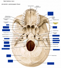
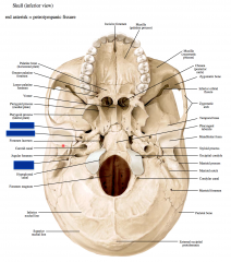
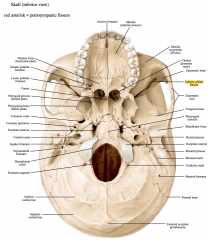
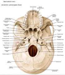
Skull: Inferior View
-Zygomatic Arch -Zygomatic process of the maxilla -Styloid process -Mastoid process -Vomer -Mastoid foramen -External occipital protuberance -Superior nuchal line -Inferior nuchal line -Occipital condyle -Mandibular fossa -Foramen magnum -Hypoglossal canal -Palatine bone -Stylomastoid foramen -Carotid canal -Pterygoid process (lateral plate) -Pterygoid process (medial plate)
|
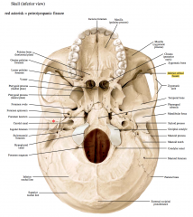
-Zygomatic Arch -Zygomatic process of the maxilla -Styloid process -Mastoid process -Vomer -Mastoid foramen -External occipital protuberance -Superior nuchal line -Inferior nuchal line -Occipital condyle -Mandibular fossa -Foramen magnum -Hypoglossal canal -Palatine bone -Stylomastoid foramen -Carotid canal -Pterygoid process (lateral plate) -Pterygoid process (medial plate) |
|
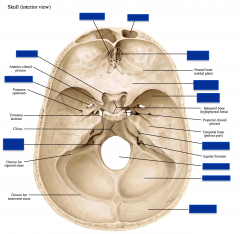
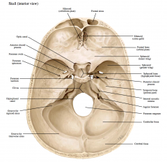
Skull: Interior View
-Foramen magnum -Cerebral fossa -Ethmoid (crista galli) -Ethmoid (cribriform plate) -Frontal sinus -Hypoglossal canal -Foramen ovale -Optic canal -Cerebellar fossa -Internal acoustic meatus -Sphenoid (lesser wing) -Sphenoid (greater wing)
|
Skull: Interior View
-Foramen magnum -Cerebral fossa -Ethmoid (crista galli) -Ethmoid (cribriform plate) -Frontal sinus -Hypoglossal canal -Foramen ovale -Optic canal -Cerebellar fossa -Internal acoustic meatus -Sphenoid (lesser wing) -Sphenoid (greater wing) |
|
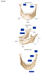
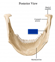
Mandible (multiple views)
-Head -Neck -Coronoid process -Mandibular foramen -Mylohyoid line |
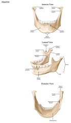
-Head -Neck -Coronoid process -Mandibular foramen -Mylohyoid line |
|
|
What are the boundaries of the parotid region, the temporal fossa, and the infra temporal fossa?
|
The following is a description of the boundaries of the following spaces:
Parotid region Superior border - zygomatic arch
Temporal fossa
Infratemporal fossa |
|
Identify the following foramina on the external surface:
-Supraorbital foramen (may be a notch) -Infraorbital foramen -Mental foramen
What bones are these foramina within?
|
Supraorbital foramen/notch – frontal bone – supraorbital nerve and vessels
Infraorbital foramen – maxillary bone – infraorbital nerve and vessels
|
|
Identify the following foramina on the external surface:
-Stylomastoid foramen -Ovale foramen -Spinosum foramen
What bones are these foramina within?
|

Stylomastoid foramen – temporal bone – facial nerve (VII) proper
Foramen spinosum – sphenoid bone – middle meningeal vessels, meningeal branch of V3 |
|

Identify the following foramina on the external surface:
-Mandibular foramen
What bones are these foramina within?
|
Mandibular foramen – mandible – inferior alveolar nerve and vessels |
|
-Inferior orbital fissure
What bones form this fissures?
What spaces does it connect?
What goes through it? |
Bones = greater wing of sphenoid bone and maxillary bone
Connects the orbit and pterygopalatine fossa
Infraorbital nerve and vessels, & zygomatic nerve run through it |
|
|
-Pterygomaxillary fissure
What bones form this fissures?
What spaces does it connect?
What goes through it? |
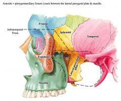
Bones = pterygoid process of sphenoid bone & maxillary bone
Connects the infratemporal and pterygopalatine fossae
Maxillary artery goes through it |
|
|
-Petrotympanic fissure
What bones form this fissures?
What spaces does it connect?
What goes through it? |
Bones = within the temporal bone (between the tympanic and petrous portions)
Connects the tympanic cavity (within the temporal bone) and infratemporal fossa
Chorda Tympani artery goes through it |
|

Superior orbital fissure:
What bones form this fissure?
What spaces does it connect?
What goes through this fissure? |
The superior orbital fissure is between the greater and lesser wings of the sphenoid bone
It connects the middle cranial fossa with the orbit
It contains the branches of the ophthalmic nerve (V1; frontal, lacrimal, nasociliary), the occulomotor nerve (III), the trochlear nerve (IV), the abducent nerve (VI) and the ophthalmic veins. |