Reading...
![]()
Play button
![]()
Play button
![]()
Use LEFT and RIGHT arrow keys to navigate between flashcards;
Use UP and DOWN arrow keys to flip the card;
H to show hint;
A reads text to speech;
110 Cards in this Set
- Front
- Back
|
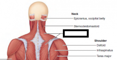
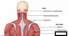
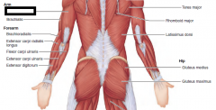
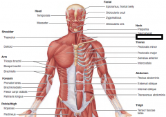
Sternocleidomastoid
|
Flexes and laterally rotates the head
origin - sternum, clavicle insertion - mastoid process of temporal |
|
|
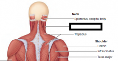
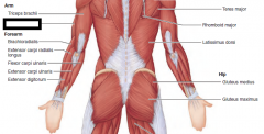
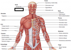
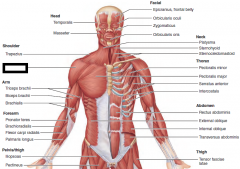
Trapezius
|
Stabilizes, raises, retracts, and rotates scapula
O—occipital bone, ligamentum nuchae, and spinous processes of C7 and all thoracic vertebrae I—a continuous insertion along acromion and spine of scapula and lateral third of clavicle |
|
|
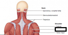
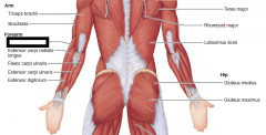
Deltoid
|
Prime mover of arm abduction when all its fibers contract simultaneously
O—embraces insertion of the trapezius; lateral third of clavicle; acromion and spine of scapula I—deltoid tuberosity of humerus |
|
|
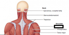
Infraspinatus
|
Rotates humerus laterally
O—infraspinous fossa of scapula I—greater tubercle of humerus posterior to insertion of supraspinatus |
|
|
Teres major
|
Extends, medially rotates, and adducts humerus;
O—posterior surface of scapula at inferior angle I—crest of lesser tubercle on anterior humerus; insertion tendon fused with that of latissimus dorsi |
|
|
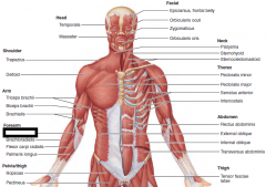
Latissimus dorsi
|
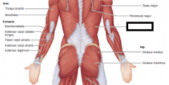
Prime mover of arm extension; powerful arm adductor; medially rotates arm at shoulder;
O—indirect attachment via lumbodorsal fascia into spines of lower six thoracic vertebrae, lumbar vertebrae, lower 3 to 4 ribs, and iliac crest; also from scapula’s inferior angle I—spirals around teres major to insert in floor of intertubercular sulcus of humerus |
|
|
Gluteus medius
|
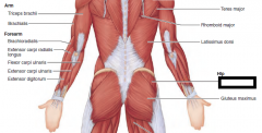
Abducts and medially rotates thigh
O—between anterior and posterior gluteal lines on lateral surface of ilium I—by short tendon into lateral aspect of greater trochanter of femur |
|
|
Gluteus maximus
|
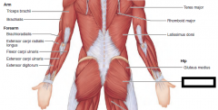
Major extensor of thigh;
origin - iliac crest, sacrum insertion - linea aspera of femur (gluteal tuberosity) |
|
|
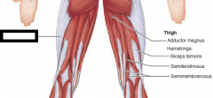
Adductor magnus
|
posterior part is a synergist of hamstrings to extend thigh
O—ischial and pubic rami and ischial tuberosity I—linea aspera and adductor tubercle of femur |
|
|
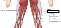
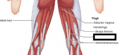
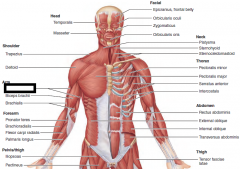
Biceps femoris
|
Extends thigh and flexes knee;
O—ischial tuberosity (long head); linea aspera, lateral supracondylar line, and distal femur (short head) I—common tendon passes downward and laterally (forming lateral border of popliteal fossa) to insert into head of fibula and lateral condyle of tibia |
|
|
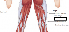
Semitendinosus
|
Extends thigh and flexes knee
O—ischial tuberosity in common with long head of biceps femoris I—medial aspect of upper tibial shaft |
|
|
Semimembranosus
|
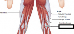
Extends thigh and flexes knee;
O—ischial tuberosity I—medial condyle of tibia; via oblique popliteal ligament to lateral condyle of femur |
|
|
Triceps brachii
|
Powerful forearm extensor
O—long head: infraglenoid tubercle of scapula; lateral head: posterior shaft of humerus; medial head: posterior humeral shaft distal to radial groove I—by common tendon into olecranon of ulna |
|
|
Brachialis
|
A major forearm flexor
O—front of distal humerus; embraces insertion of deltoid muscle I—coronoid process of ulna and capsule of elbow joint |
|
|
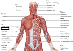
Brachioradialis
|
Synergist in flexing forearm;
O—lateral supracondylar ridge at distal end of humerus I—base of radial styloid process |
|
|
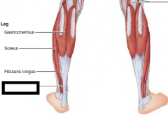
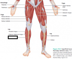
Iliotibial tract
|
d
|
|
|
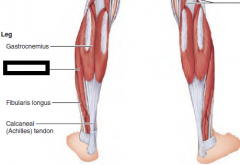
Gastrocnemius
|
Plantar flexes foot
O—by two heads from medial and lateral condyles of femur I—posterior calcaneus via calcaneal tendon |
|
|
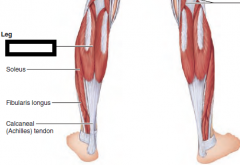
Soleus
|
Plantar flexes foot
O—extensive origin from superior tibia, fibula, and interosseous membrane I—as for gastrocnemius |
|
|
Calcaneal tendon
|
(Achilles)
|
|
|
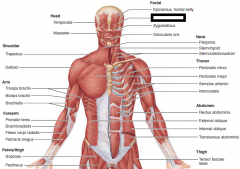
Temporalis
|
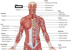
O—temporal fossa
I—coronoid process of mandible via a tendon that passes deep to zygomatic arch Closes jaw; |
|
|
Masseter
|
O—zygomatic arch and zygomatic bone
I—angle and ramus of mandible Prime mover of jaw closure; elevates mandible |
|
|
Trapezius
|

O—occipital bone, ligamentum nuchae,
and spinous processes of C7 and all thoracic vertebrae I—a continuous insertion along acromion and spine of scapula and lateral third of clavicle Stabilizes, raises, retracts, and rotates scapula |
|
|
Deltoid
|
O—embraces insertion of the trapezius;
lateral third of clavicle; acromion and spine of scapula I—deltoid tuberosity of humerus Prime mover of arm abduction when all its fibers contract simultaneously; |
|
|
Triceps brachii
|
O—long head: infraglenoid tubercle
of scapula; lateral head: posterior shaft of humerus; medial head: posterior humeral shaft distal to radial groove I—by common tendon into olecranon of ulna Powerful forearm extensor |
|
|
Biceps brachii
|
origin - glenoid fossa, corocoid process of scapula
insertion - radial tuberosity |
|
|
Brachialis
|
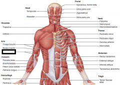
O—front of distal humerus; embraces
insertion of deltoid muscle I—coronoid process of ulna and capsule of elbow joint A major forearm flexor |
|
|
Pronator teres
|
O—medial epicondyle of humerus; coronoid process of ulna
I—by common tendon into lateral radius, midshaft Pronates forearm; weak flexor of elbow |
|
|
Brachioradialis
|
arises from the distal humerus and inserts
on the distal forearm, it resides mainly in the forearm. Its force is exerted far from the fulcrum, so the brachioradialis is a weak forearm flexor |
|
|
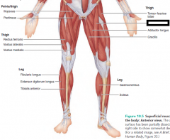
Iliopsoas
|
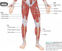
Iliopsoas is a composite of two closely related muscles (iliacus and psoas major) whose fibers pass under the
inguinal ligament (see Figure 10.12) to insert via a common tendon on the femur. |
|
|
Pectineus
|
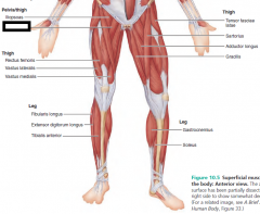
O—pubis (and superior
ramus) I—from lesser trochanter inferior to the linea aspera on posterior aspect of femur Adducts, flexes, and medially rotates thigh |
|
|
Rectus femoris
|
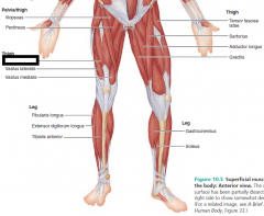
origin - anterior inferior iliac spine
insertion - proximal anterior surface of tibia Extends knee and flexes thigh at hip |
|
|
Vastus lateralis
|
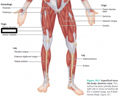
origin - linea aspera of femur
insertion - proximal anterior surface of tibia Extends and stabilizes knee |
|
|
Vastus medialis
|
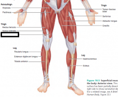
origin - linea aspera of femur
insertion - proximal anterior surface of tibia Extends knee |
|
|
Tibialis anterior
|
O—lateral condyle and
upper 2/3 of tibial shaft; interosseous membrane I—by tendon into inferior surface of medial cuneiform and first metatarsal bone Prime mover of dorsiflexion; inverts foot; helps support medial longitudinal arch of foot |
|
|
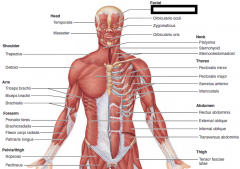
Epicranius, frontal belly
|
|
|
|
Orbicularis oculi
|
|
|
|
Zygomaticus
|
|
|
|
Orbicularis oris
|
|
|
|
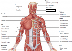
Sternocleidomastoid
|
|
|
|
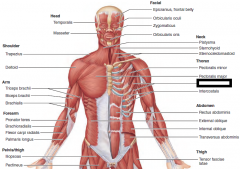
Pectoralis major
|

origin - clavical, sternum
insertion - greater tubercle of humerus |
|
|
Serratus anterior
|
O—by a series of muscle
slips from ribs 1–8 (or 9) I—entire anterior surface of vertebral border of scapula Rotates scapula so its inferior angle moves laterally and upward; |
|
|
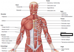
Rectus abdominis
|
|
|
|
External oblique
|
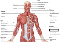
|
|
|
Internal oblique
|
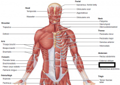
|
|
|
Transversus abdominis
|
|
|
|
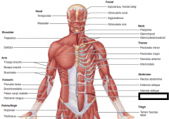
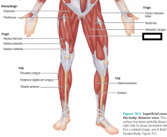
Tensor fasciae latae
|
O—anterior aspect of
iliac crest and anterior superior iliac spine I—iliotibial tract* Steadies the knee and trunk on thigh by making iliotibial tract taut; flexes and abducts thigh; rotates thigh medially |
|
|
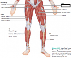
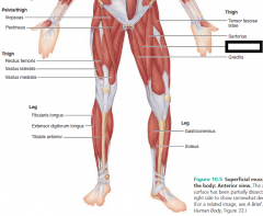
Sartorius
|
origin - anterior superior iliac spine
insertion - medial surface tibia Flexes, abducts, and laterally rotates thigh; flexes knee (weak) as in a soccer kick; helps produce the cross-legged position |
|
|
Adductor longus
|
O—pubis near pubic
symphysis I—linea aspera Adducts, flexes, and medially rotates thigh |
|
|
Gracilis
|
O—inferior ramus and
body of pubis and adjacent ischial ramus I—medial surface of tibia just inferior to its medial condyle Adducts thigh, flexes and medially rotates leg, especially during walking |
|
|
Gastrocnemius
|
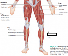
origin - medial and lateral condyles of femur
insertion - calcaneous via Achilles tendon Plantar flexes foot when knee is extended; because it also crosses knee joint, it can flex knee when foot is dorsiflexed |
|
|
Soleus
|
O—extensive origin from
superior tibia, fibula, and interosseous membrane I—as for gastrocnemius Plantar flexes foot; important locomotor and postural muscle during walking, running, and dancing |
|
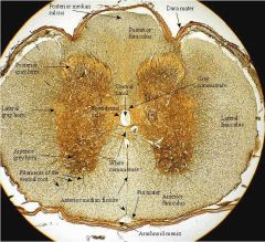
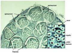
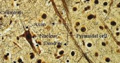
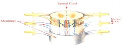
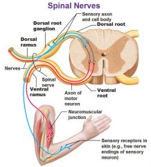
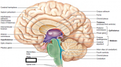
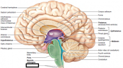
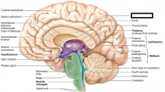
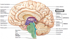
White matter,
gray matter, posterior horn, anterior horn, central canal, sensory root, sensory root ganglion (sensory neuron cell body, nucleus), motor root, motor neurons |
|
|
cell
location parts |
|
|
cell
location parts |
|
|
cell
location parts |
|
|
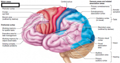
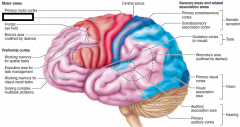
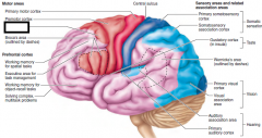
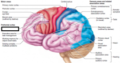
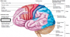
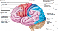
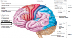
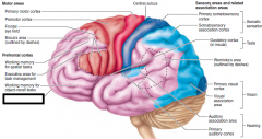
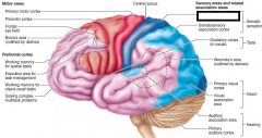
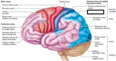
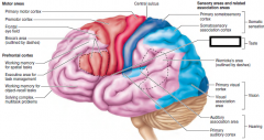
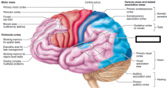
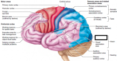
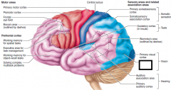
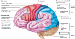
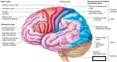
motor area?
|
|
|
|
primary motor area
|
|
|
|
premotor cortex (planning)
|
|
|
|
Frontal eye field
|
|
|
|
prefrontal corted
working memory for spatial tasks |
|
|
|
prefrontal
executive area for task management |
|
|
|
broca's area
|
|
|
|
working memory for object recall tasks
|
|
|
|
solving complex, multitask problems
|
|
|
|
Primary somatosensory cortex
|
|
|
|
somatosensory association area
|
|
|
|
gustatory cortex
|
|
|
|
Wernicke's area
|
|
|
|
primary visual cortex
|
|
|
|
visual association area
|
|
|
|
auditory association area
|
|
|
|
primary auditory cortex
|
|
|
|
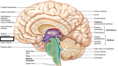
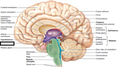
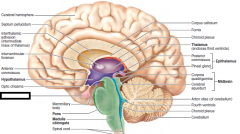
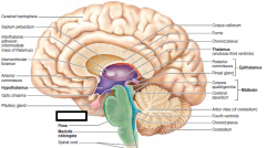
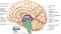
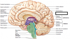
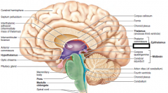
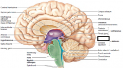
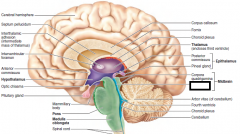
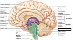
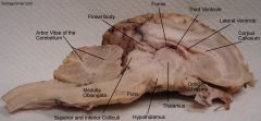
Hypothalamus
|
|
|
|
optic chiasma
|
|
|
|
pituitary gland
|
|
|
|
mammillary body
|
|
|
|
pons
|
|
|
|
medulla oblongata
|
|
|
|
spinal cord
|
|
|
|
corpus callosum
|
|
|
|
choroid plexus
|
|
|
|
thalamus(encloses third ventricle)
|
|
|
|
pineal gland
|
|
|
|
corpora quadrigemina
|
|
|
|
cerebral aqueduct
|
|
|
|
arbor vitae
|
|
|
|
fourth ventricle
|
|
|
|
choroid plexus
|

|
|
|
cerebellum
|
|
|
|
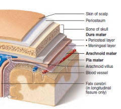
Epidural Space
Subdural Space Subarachnoid Space |
|
|
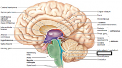
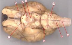
Frontal lobe of cerebellum
Optic chiasma parietal lobe cerebellum pineal gland arbor vitae corpora quadrigemina fourth ventricle medulla oblongata pons |
|
|
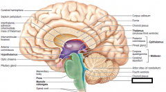
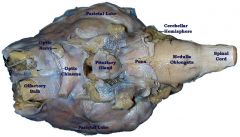
Olfactory bulb
Optic nerve Pituitary/Mammillary Body Pons Medulla oblongata |
|
|
|
I. Olfactory
|
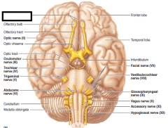
Purely sensory-carries impulses for sense of
smell . Person is asked to sniff and identify aromatic substances, such as oil of cloves and vanilla |
|
|
II. Optic
|
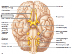
Vision and visual field are determined with eye
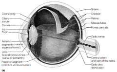
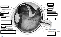
chart and by testing the point at which the person first sees an object (finger) moving into the visual field. Eye interior viewed with ophthalmoscope to detect swelling of optic disc (point at which optic nerve leaves the eye) and to observe blood vessels. |
|
|
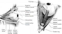
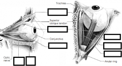
III. Oculomotor
|
Mixed-motor fibers to inferior oblique and
superior, inferior, and medial rectus muscles, which direct eyeball; to levator palpebrae muscles of eyelid; to iris and smooth muscle controlling lens shape and pupil size. Pupils are examined for size, shape, and equality. Pupillary reflex is tested with penlight (pupils should constrict when illuminated). Convergence for near vision is tested, as is subject's ability to follow objects up, down, side to side, and diagonally with eyes. |
|
|
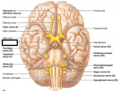
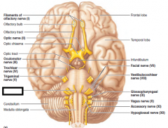
IV. Trochlear
|
Mixed-provides motor fibers to superior
oblique muscle (an extrinsic eye muscle). Tested in common with cranial nerve III. |
|
|
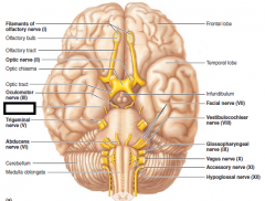
V. Trigeminal
|
Mixed-conducts sensory impulses from skin
of face and anterior scalp, from mucosae of mouth and nose. Also contains motor fibers that activate the chewing muscles. Sensations of pain, touch, and temperature are tested with safety pin and hot and cold objects. Corneal reflex tested with wisp of cotton. Motor branch assessed by asking person to clench his teeth, open mouth against resistance, and move jaw side to side. |
|
|
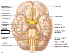
VI. abducens
|
Carries motor fibers to lateral rectus muscle
of eye. Tested in common with cranial nerve III. |
|
|
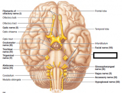
VII. facial
|
Mixed-supplies motor fibers to muscles of
facial expression and to lacrimal and salivary glands; carries sensory fibers from taste receptors of anterior tongue. Anterior two-thirds of tongue is tested for ability to taste sweet (sugar), salty, sour (vinegar), and bitter (quinine) substances. Symmetry of face is checked. Subject is asked to close eyes, smile, whistle, and so on. Tearing is assessed with ammonia fumes. |
|
|
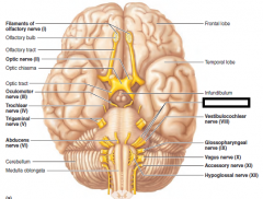
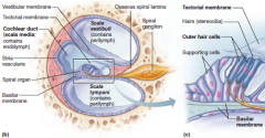
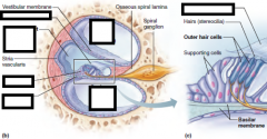
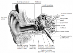
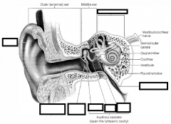
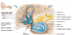
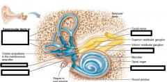
VIII. vestibulocochlear
|
Purely sensory-transmits impulses for senses
of equilibrum and hearing. Hearing is checked by air and bone conduction using tuning fork. |
|
|
IX. glossopharyngeal
|
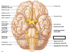
Mixed-motor fibers serve pharyngeal
muscles and salivary glands; sensory fibers carry impulses from pharynx, posterior tongue (taste buds), and pressure receptors of carotid artery. Gag and swallowing reflexes are checked. Subject is asked to speak and cough. Posterior third of tongue may be tested for taste. |
|
|
X. vagus
|
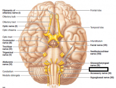
Mixed-Motor fibers to pharynx and larynx
and sensory fibers from same structures; a very large portion is composed of parasympathetic motor fibers, which supply heart and smooth muscles of abdominal visceral organs; transmits sensory impulses from viscera. As for cranial nerve IX (IX and X are tested in common, because they both serve muscles of throat and mouth). |
|
|
XI accessory
|
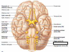
Mixed-provides motor fibers to
sternocleidomastoid and trapezius muscles. Sterncleidomastoid and trapezius muscles are checked for strength by asking person to rotate head and elevate shoulders against resistance. |
|
|
XII. hypoglossal
|
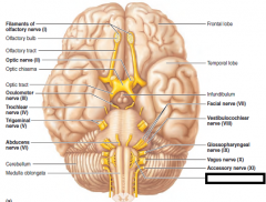
M ixed-motor fibers serve muscles of tongue
and sensory fibers carry impulses from tongue. Person is asked to protrude and retract tongue. Any deviations in position are noted. |
|
|
|
|
|
|
|
|
|
|
|
|
|
|
|
|
|
|