Reading...
![]()
Play button
![]()
Play button
![]()
Use LEFT and RIGHT arrow keys to navigate between flashcards;
Use UP and DOWN arrow keys to flip the card;
H to show hint;
A reads text to speech;
83 Cards in this Set
- Front
- Back
|
What is the Vertebral formula = # of vertebrae in each region for cat and dog?
|
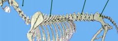
For dog & cat = C7, T13, L7, S3, Cd ≈ 20
|
|
|
What is Shiff-Sherrington syndrome?
|
Hyperextension of forelimbs with damage/lesions to thoracic spinal cord (T3-L3 / area 3)
Only time a spinal cord injury causes signs cranial to damage/injury LMN and UMN are still intact to thoracic limb, so this is neither UMN or LMN neuron sign. (hindlimb may show UMN signs) From Online: Extensor hypertonia (increased muscle tone ) of the thoracic limbs and paraplegia resulting from acute, severe, compressive lesions of the thoracolumbar spinal cord that REMOVE the inhibitory effects of neurons in the lumbar spinal cord. Seen in dogs, usually caused by trauma or herniated intervertebral disk. Border cells in L1-4 ascend to inhibit extensors of forelimb to coordinate movement |
|
|
What is the main hypaxial/sublumbar muscle of the abdomen?
|
Psoas major m.
hypaxial is below transverse processes of spine |
|
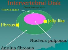
Intervertebral Disk or fibrocartilaginous disks consists of inner jelly like part and outer part known as the _____ and ______?
|

nucleus pulposus
anulus fibrosus |
|
|
for tards:
difference between vertebral column and spinal cord |
- vertebrae are actual bone
- protect the spinal cord |
|
|
Intervertebral Disk or fibrocartilaginous disks consists of inner jelly like part and outer part known as the _____ and ______?
|
nucleus pulposus
anulus fibrosus |
|
|
Intrathecal injection
|

in Lumbar cistern- between L4-5 or L5-6
access to CSF (subarachnoid space) |
|
|
Epidural anesthesia goes where?
|
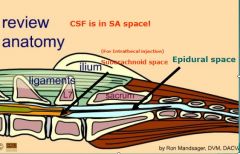
lumbosacral junction (in small animals) in epidural space
Epidural space: between the dura mater & vertebrae Filled with fat Epidural anesthesia is performed within this space |
|
|
Name 3 epaxial muscles
|

Epaxial Muscle Systems = “TLI”
transversospinalis muscles (4) longissiumus muscles (4) iliocostalis muscles (2) the later two break up into muscles based on region of spinal cord e.g. Iliocostalis lumborum |
|
|
List the spaces that are related to the meninges
|
* Epidural: between dura mater and periosteum
* Subdural: potential space between the dura mater and arachnoid * Subarachnoid space: between the arachnoid and pia (CSF) |
|
|
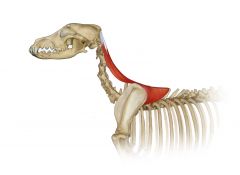
What is the main HYPAXIAL muscle of the CERVICAL region?
|
Longus colli
|
|
|
What is the disease of the cervical vertebrae in large breeds causing STENOSIS of the vertebral canal resulting in ataxia (unsteady gait)?
|
Cervical spondylomyelopathy, canine wobbler disease
|
|
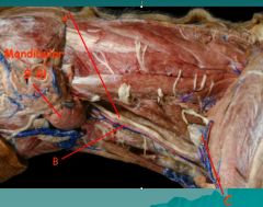
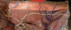
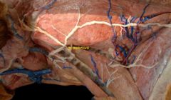
Identify
|
A. Vagosympathetic trunk
B. Common carotid a. C. Supf. Cervical Ln |
|
|
What is excessive ventral lumbar curvature?
|
Lordosis (swayback)
|
|
|
What is spina bifida?
|
Failure of 1 or more vertebral arches to close
|
|
|
A slipped disc results when the soft ___ ___ is squeezed to one side of the disc, causing the firm ___ ___ to protrude and possibly rupture.
|
* Nucleus pulposus
* Annulus fibrosus |
|
|
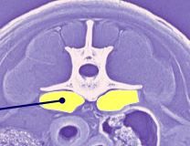
Where are CSF taps performed?
~looking for name of region or place, not the layer |
Cisterna magna or lumbar cistern (subarachnoid space)
|
|
|
What kind of joint is intervertebral disk?
|

Symphysis (cartilagenous)(e.g., pelvis, mandible, intervertebral disk) - semimobile
~but i think disk itself is made of fibrocartilage |
|
|
What is the lumbosacral space?
|

Interarcuate space between last lumbar vertebrae (L7) & sacrum
(where insert needle when giving epidural injection) |
|
|
anticlinal vertebrae
|
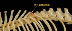
T11
|
|
|
How is proprioception evaluated clinically?
|
Postural reactions (e.g. knuckle paw)
|
|
|
how many lumbar vertebrae in cat and dog?
|
7
|
|
|
identify thoracolumbar spinal cord
(what is located here?) |
Of or relating to the thoracic AND lumbar parts of the spinal column.
sympathetic division of the autonomic nervous system (specifically thoracolumbar: T3 to L3) |
|
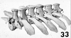
identify which region of vertebrae this is?
|
Lumbar Vertebrae
Elongated transverse processes Relatively mobile, particularly in extension-flexion movements 7 in dog & cat |
|
|
What arises from spinal cord to FORM a spinal nerve?
What arises from spinal nerve? |
dorsal and ventral roots (NOT branches)
dorsal and ventral branches; these are formed when spinal nerve "branches" |
|
|
hemal bones (arches)
|
Any of three or four V-shaped bones located ventral to the bodies of the third to sixth CERVICAL vertebrae
|
|
|
Are spinal nerves LMN or UMN?
|
LMN (somatic)
|
|
Identify
|
intercapital liigament
|
|
|
Where are the cell bodies of a UMN and LMN located?
What are axons of upper and lower motor neurons? |
in brain and spinal cord respectively
UMN: brain and spinal c. LMN: periphery |
|
|
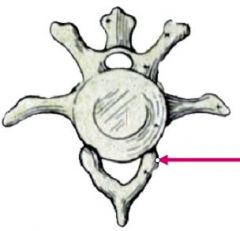
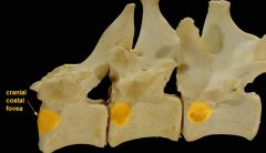
Where does tubercle of ribs articulate with on vertebrae?
|
transverse process
|
|
|
What are the motor parts of reflex arcs?
|
LMN
|
|
|
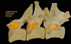
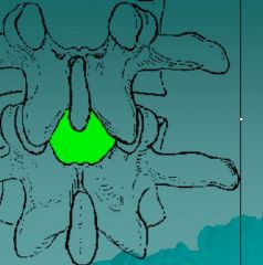
Where does head of rib articulate with on vertebrae?
|
see image
|
|
|
Is musculocutaneous nerve an upper or lower motor neuron?
|
LMN
|
|
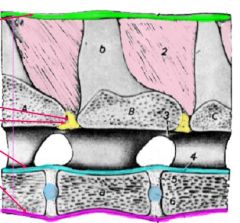
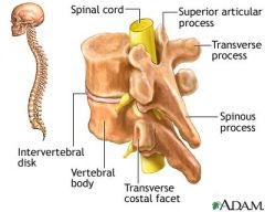
Identify 4 ligaments shown
|
Supraspinous lig.
Yellow lig. Dorsal longitudinal lig. Ventral longitudinal lig. |
|
|
What is the enlargement of the subarachnoid space between the medulla oblongata and cerebellum?
|
Cisterna magna (cerebellomedullary cistern)
|
|
|
lig. flava is also known as?
|
Yellow ligament - connect successive vertebral arches
|
|
|
The ventral branches of the thoracic nerves T3-13 do not form a plexus, but pass in the intercostal spaces as _____ nerves.
|
intercostal
|
|
|
Where do the spinal nerves leave the vertebral canal?
|
intervertebral foramen
|
|
|
What are the clinical signs of meningitis?
|
Cervical rigidity (due to pain [hyperesthesia]), fever and lameness
Hyperesthesia - abnormal increase in sensitivity to stimuli of the senses. |
|
|
Name the two major epaxial muscles of the back
|
Iliocostalis and Longissimus mm.
|
|
|
What does the sacral region of the spinal cord supply?
|
Reflex control of urination, defication, sexual reflexes, and parasympathetic outflow
|
|
|
What is a dermatome? Autonomous zone?
|
Area of skin innervated by a nerve, only 1 spinal nerve respectively
|
|
|
List the meninges from outer to inner
|
Dura matter, arachnoid, and pia mater
1. Dura mater- “tough mother” - outermost layer (epidural space is above/outside of this) 2. Arachnoid- web-like layer; next to dura mater in living because of CSF pressure Subarachnoid space—FILLED WITH CSF!!! 3. Pia mater- “tender mother” layer on surface of cord, contains blood vessels supplying brain and cord tissue |
|
|
What fills the gap between the dorsal edge of the foramen magnum and the atlas?
|
Dorsal atlanto-occipital membrane
(this is what needle goes through to get to cisterna magna - for CSF) p.315 |
|
|
What is the UNPAIRED artery running longitudinally on the vertebral canal floor in the ventral median fissure length of the spinal cord
|
Ventral spinal artery
(runs longitudinally inside bottom of vertebral foramen, beneath spinal cord, but on top of body of vertebrae) |
|
|
What are the PAIRED, thin- walled, valveless vessels on the vertebral canal floor in the epidural space from the skull to the caudal vertebrae?
|
Internal vertebral venous plexus
|
|
|
What are the vessels located on the ventral surface of the tail?
|
Median caudal artery and vein
|
|
|
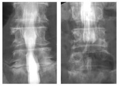
How are the dorsal and ventral edges of the vertebral canal checked in back radiographs?
|
Check for alignment, they should be two straight lines without step defects
|
|
|
How are the intervertebral foramen compared in back radiographs?
|
For differences due to disc space differences
|
|
|
Myelography
|
Type of radiograph exam that uses contrast medium to detect pathology of spinal cord, including location of injury, cysts, and tumors. Procedure often involves injection of contrast medium into cervical or lumbar spine, followed by several X-rays. May help to find cause of pain not found by an MRI or CT.
|
|
|
What may narrowing of the intervertebral space indicate?
|
Protruded disc
|
|
|
Describe the three possible types of spinal column lesion
|
* Extradural lesion: outside the dura mater
* Intradural lesion: between spinal cord and dura mater * Intramedullary (spinal cord) lesion: inside cord |
|
|
What are the myelogram findings for the following lesions:
Extradural, intradural, and intramedullary (spinal cord) lesions |
* Extradural: thinning or break of columns pushed inward at lesion, expanded cord/ thinned columns in other view
* Intradural: widening of subarachnoid space, expanded cord/ thinned columns in other view * Intramedullary: Expanded cord/ thinned columns in all views |
|
|
What is lateral curvature to the spine?
|
Scoliosis
|
|
|
What is an excessive thoracic curvature?
|
Kyphosis (hunchback in humans)
|
|
|
What clinical problem would a fractured dens cause?
|
spinal cord injury
|
|
|
What is a hemivertebra?
|
Wedge-shaped vertebra resulting in severe angulation to spine in thoracolumbar region.
|
|
|
What is diskospondylitis?
|
Infection of intervertebral discs & adjacent vertebrae
|
|
|
Basically what are the two types of intervertebral disease?
|
* Type 1: disk rupture
* Type 2: disk bulging |
|
|
What can a slipped disc protruding into the vertebral canal compress?
|
Spinal nerves or spinal cord itself
|
|
|
Where is the annulus fibrosis thinnest?
|
Dorsally
|
|
|
Why don't intervertebral disc commonly impinge on nerves in most of the thoracic region?
|
Protection of intercapital ligaments
|
|
|
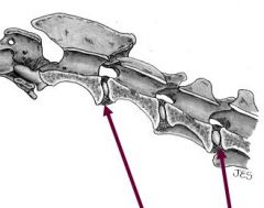
Where is rupture of an intervertebral disc common?
|
# Thoracolumbar junction (T11-L2)
|
|
|
What is the surgery for thoracolumbar disk disease?
|
Dorsal laminectomy/hemilaminectomy & fenestration (remove nucleus pulposus)
|
|
|
What are the landmarks used for cerebrospinal fluid taps at the atlanto-occipital junction?
|
Wings of the atlas, spine of the axis, external occipital protuberance
|
|
|
Largest group of of sesamoid bones of manus is located at lvl of the____________ joint.
|
metacarpophalangeal joint
|
|
|
What is path of LMN?
|
Leave CNS over VENTRAL roots (only one motor neuron) & spinal n. or cranial n. to periphery
|
|
Deep to the trapezius and brachiocephalicus, a pair of muscles called __________ arise from the dorsal midline (um no back of the head) and insert on the proximal end of each scapula near dorsal midline (um no back of the head) and insert on the proximal end of each scapula near dorsal border.
|
rhomoideus
(actually it kind of drapers over top of scapula from back of the head) |
|
|
What type of joint is atlanto-occipital joint?
|
- modified-hinge joint; synovial
- formed between condyles of occipital bone and atlas "yes joint" |
|
|
What type of joint is atlanto-axial joint?
|
pivot type of synovial joint between dens of axis? and atlas
"no joint" dens is held into place with a lot of ligaments |
|
|
ataxia
plegia |
unsteady gait
partial paralysis |
|
|
What joint is formed by the atlas and the skull?
|
Atlanto-occipital joint, "yes" joint
|
|
|
What elastic connective tissue structure attaches the 1st thoracic spine to the spine of the axis (C2) in the dog?
|
Nuchal ligament, none in cat
|
|
|
What plexus supplies some of the extrinsic and all of the intrinisic muscles of the thoracic limb?
|
Brachial
|
|
|
Plexuses are formed by the ventral branches of spinal nerves in every region except which?
|
Thorax (except T1-2)= intercostal nn.
|
|
|
What plexus supplies the abdominal wall, pelvic limb, external genitalia, rump, and perineum?
|
Lumbrosacral plexus
|
|
|
Why can a broken neck result in respiratoy paralysis?
|
Phrenic nerve to diaphragm arises in brachial & cervical plexuses
|
|
|
What should be evaluated in the area of the axis and atlas?
|
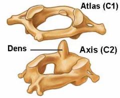
The dens (odontoid process), it should be present and held in the ventral veterbral canal
odontoid means "looks like a tooth" odontoid p. is a toothlike projection on the cranial part of body of the axis and articulates with the atlas |
|
|
What is a landmark in a lateral film of the caudal neck?
|
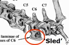
"SLEDS" or transverse processes of C6
|
|
|
List the two important structures enclosed in the carotid sheath.
|
vagosympathetic trunk and common carotid artery
|
|
|
What is the surgery for cervical disc disease?
|
Ventral decompression through longus colli m. to remove extruded disk
|
|
What nerve is this?
(hint: it innervates trapezius muscle which is transfected in image) |
accessory nerve (cranial n. 11)
|
|
|
Name strap muscle (plus muscle lateral to them), starting at midline
|
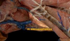
sternohyoideus m.,, sternothyroideus m.,
sternocephalicus m. cleidocephalicus m. |