Reading...
![]()
Play button
![]()
Play button
![]()
Use LEFT and RIGHT arrow keys to navigate between flashcards;
Use UP and DOWN arrow keys to flip the card;
H to show hint;
A reads text to speech;
155 Cards in this Set
- Front
- Back
|
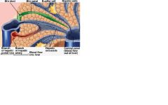
Describe how the liver obtains oxygenated blood
|
Hepatic artery branches off the abdominal aorta and enters the hilus of the liver
|
|
|
List the vessels of the liver hilus
|
Hepatic artery, portal vein, common hepatic duct
|
|
|
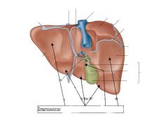
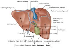
Looking from the rear of the liver, describe the 4 lobes
|
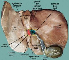
1) Left lobe left of ligamentum venosum and ligamentum teres
2) Caudate lobe superior to hilus, right of ligamentum venosum and left of bare area. 3) Quadrate lobe right of ligamentum teres and inferior to hilus, left of gall bladder 4) Right lobe: right of caudate lobe, hilus and gall bladder and includes bare area. |
|
|
List the vessels on the 6 corners and centre of an hepatic lobule
|
1) portal vein
2) hepatic artery 3) bile duct 4) central: central vein -> hepatic vein |
|
|
Describe the ligaments of the bare area and their relationship to the peritonia
|
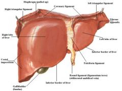
1) The coronary ligaments reflect the visceral peritonium upward against the diaghram and become the parietal peritonium.
2) The anterior leaves of the coronary ligaments become the falciform ligament 3) The posterior leaves become the lesser omentum. |
|
|
List the structures housed within the thoracic cavity
|
2xpleural cavities, mediastinum contaning the heart, thymus, trachea, bronchi, lungs
|
|
|
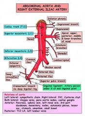
List the 3 vessels branching off the coeliac trunk
|
1) splenic (left)
2) left gastric (up) 3) common hepatic (right) |
|
|
What is the 1st artery branching off the common hepatic artery
|
Gastroduodenal
|
|
|
What are the 2 arteries branching from the gastroduodenal
|
1) superior pancreaticoduodenal
2) right gastro-omental |
|
|
What branches off the hepatic arteries
|
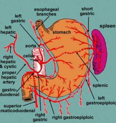
1) right gastric
2) gastroduodenal 3) superior pancreaticoduodenal |
|
|
Describe the 3 layers of fasciae
|
1) superficial within the reticular layer of the dermis
2) deep: surrounds muscles, nerves, bones, blood vessels 3) visceral: suspends organs within their cavities eg fibrous pericardium |
|
|
How can oedema deep within the thigh lead to tissue necrosis
|
Oedema -> increased pressure due to unyielding fascia surrounding muscles -> constriction of blood vessels -> tissue necrosis
|
|
|
Which parts of the skeleton does the pelvic girdle belong to
|
Appendicular: ilium, ischium, pubis belong to the appendicular skeleton.
Axial: sacrum, sacraliliac joint |
|
|
Describe the formation and development of the embryonic disk
|
1) Cells of cytotrophoblast distal to uterine cavity proliferate into amnion & space
2) Amnion towards centre of egg forms endoderm (next to yolk) and ectoderm (the embryonic disk) 3) Endoderm surrounds yolk sac, mesoderm forms and ectoderm surrounds amnionic space. 3 layers form the embryonic disk. |
|
|
Describe the general differentiation into structures by the germ layers
|
Endoderm: epithelial lining of GI, respiratory tract, liver, GB, pancreas.
Mesoderm: muscle, bones, connective tissue, endothelium Ectoderm: epidermis & nervous system |
|
|
List the locations of the enteric nerve plexi within the GI wall
|
1) myenteric plexus between circular & longtitudinal layer
2) submucosal plexis in the submucosa |
|
|
List the layers of the GI tract
|
1) serosa
2) Muscularis: longtitudinal, circular 3) submucosa 4) mucosa (muscularis mucosa, lamina propria, mucous epithelium) |
|
|
What are the 3 levels of control of motility & secretions of the GIT
|
1) Neural
2) endocrine (gastrin, secretin, VIP, CCK, GIP, motilin) 3) Paracrine (histamine) |
|
|
Describe how sensory information from the GIT is acted on by the nervous systems
|
1) parasympathetic afferents: mainly mechanoreceptors
2) sympathetic (splanchic) afferents: chemoreceptors both within dorsal root ganglia 3) local afferents (sensory, secretory, mechanical) All lead to respective efferents: smooth muscle coordination, secretions, hormones, vasodilation/constriction |
|
|
From medial to lateral, list the order of the most major vein, artery & nerve in the femoral triangle
|
VAN - femoral vein, artery nerve
|
|
|
Adduction
|
Flaps like a duck ie brings arms closes to sides (saggital plane)
|
|
|
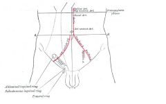
List the boundaries of the femoral triangle relative to the centre
|
superior: inguinal ligament
medial:adductor longus lateral: sartorius |
|
|
Where is the rectus femoris relative to the femoral triangle
|
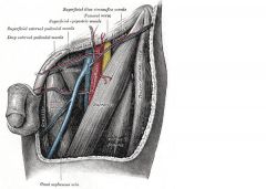
Lateral to the sartorius
|
|
|
Where is the gracilis relative to the femoral triangle
|
Medial to the rector femoris
|
|
|
What is posterior and anterior to the femoral triangle
|

posterior: iliacus (lateral), pectineus (medial)
anterior: fascia lata and sartorius just beneath that |
|
|
Out of the aorta, pulmonary trunk, pulmonary veins, inferior vena cava, which carry oxygen
|
aorta, pulmonary veins
|
|
|
How is the femoral artery positioned relative to the femoral psoas muscles
|
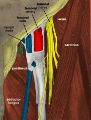
iliacus (lateral), pectineus (medial)
|
|
|
List the 5 forms of shock
|
1) cardiogenic
2) hypovolemic 3) septic 4) anaphylactic 5) neurogenic |
|
|
List the 3 main branches and sub-branches of the aortic arch
|
Moving medial to lateral (right to left)
1) brachiocephalic (right common carotid, right subclavian) 2) left common carotid 3) left subclavian |
|
|
Describe the carotid sinus
|
1) junction of the common carotid, internal & external carotids
2) has high pressure baroreceptors |
|
List the labellelled regions
|
How many correct?
|
|
|
List the blood vessels
|
How many?
|
|
Name the blood vessels
|
How many did you get
|
|
|
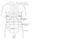
Define the subcostal plane
|
Beneath the lowest point of the ribcage (1th rib) = top of L3
|
|
|
Define the plane of the iliac crests
|
Top of iliacs = top of T4
|
|
|
Define the costal margin
|
From the 7th costal cartilage at the xyphoid to the tip of the 12th rib
|
|
|
From T9 down, define the vertebral surface landmarks
|
T9: xyphoid
L1: transpyloric plane 1/2 way between the suprasternal notch & pubic symphysis L3: subcostal margin beneath 10th rib L4: posterior tops of illiac crests L5: tubercles of the iliac crests |
|
|
Define the transpyloric plane and how it is a surfave marking for several organs
|
Transpyloric plane = mid-way between suprasternal notch and pubic symphysis
1) duodenal flexure 2) pyloris 3) pancreatic neck 4) gallbladder fundus 5) hilus of the kidneys |
|
|
Describe the surface markings of the liver
|
5-5-10 ie 5th rib on each side, 10th rib on right
|
|
|
Describe the abdominal aorta according to surface markings
|
Behind liver, slightly left of centre, bifurcating at plane of iliac creasts T4
|
|
|
Describe the attachment of the diaphragm to the surface markings of the rib cage
|
Joins at the xifisternal junction and goes up to the 5th rib on left & 4th rib on right around the nipple, then drops down to the bottom of the costal margin
|
|
|
How do we landmark the vertical lateral lines of the abdomen
|
At the mid point between the anterior superior iliac crest and the umbilicus
|
|
|
Using surface markings, define the pubic cavity
|
Bottom of L5, defined by the iliac spines down to the pubic symphysis
|
|
|
Describe the surface landmarks of the bifurcation of the abdominal aorta
|
Bifurcates at iliac creasts (L4)
|
|
|
Describe the vertebral lebel of the sternal angle and the important relations
|
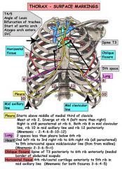
Left base of the heart
Start of aortic arch Bifurcation of trachea |
|
|
Describe the surface anatomy of the inferior vena cava
|
Protrudes the diaphragm at T8, down along the right of the abdominal aorta and down to L5 and bifurcates. Both iliac veins are medial to the iliac arteries and the IVC passes behind the right iliac artery
|
|
|
Describe the surface markings of the kidneys
|
The lower pole at L3, Upper pole at T12, hilus at the transpyloric plane. The lateral margins also pass through the hilus. The right kidney is about 2cm below the left due to the liver. The suprarenal glands form a crescent on the left and a cone on the right, tucked behind the vena cava.
|
|
|
Describe the surface markings of the duodenum
|
Begins to the left of the vena cava at the trans pyloric plane, down to the bottom poles of the kidneys, crosses in front of the aorta and vena cava, and back up to L2.
|
|
|
Describe the surface markings of the spleen
|
Above the transpyloric plane just beneath the diaphragm, just lateral to the lateral line and located posteriorly
|
|
|
Describe the surface markings of the spleen
|
Head encircled by the duodenum, tail ends at the centre of the hylum of the spleen.
|
|
|
How is does the stomach and duodenum differ in mucous secretion
|
Stomach: epithelial mucosa and neck mucosal cells secrete directly into the lumen
duodenum: goblet cells within the submucosa secrete into ducts |
|
|
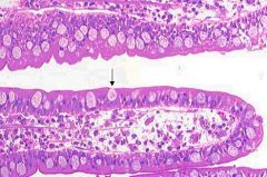
Compare the luminal surface anatomy and glands of the duodenum, small intestine and colon
|
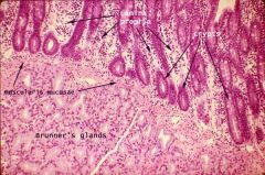
duodenum: villi and crypts, submucosal Brunner's and goblet cells lining villi and crypts
SI: villi & crypts, goblet cells lining villi and crypts Colon: crypts, goblet cells lining surface and; crypts |
|
Signs of neonatal hypoglycemia
|
-Jitteriness -Poor muscle tone -Diaphoresis
-Poor suck -Tachypnea -Dyspnea -Cyanosis -Apnea -Low temp -High-pitched cry -Irritability -Lethargy -Seizures, coma -Some infants may be asymptomatic |
|
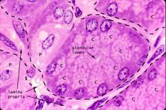
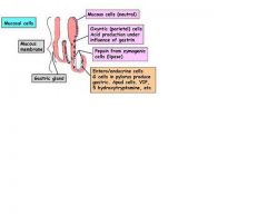
Describe P, C, and their location
|
Parietal & chief cells of the gastric glands within the mucosa
|
|
to gather, collect
|

جَمَعَ
يجمع الجمع |
|
|
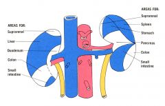
List the anterior, posterior, superior, lateral left, lateral right relations of the pancreas
|
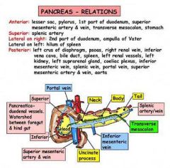
anterior: 1st part of duodenum, lesser sac, stomach, transverse mesocolon
posterior: psoas, spenic artery & vein, left kidney & vessels, superior mesenteric artery & vein, portal vein, inferior vena cava aorta left lateral: spleen right lateral:: part 2 of duodenum, |
|
|
Layers of the stomach
|
Outer longtitudinal
Inner circular Innermost oblique |
|

Name the structures of the gastric gland
|
Make sure you know why each is where it is
|
|
Name the structures
|
Make sure you know why each is where it is
|
|
|
Ligaments: list the ones joining the femur to the hip
|
pubofemoral, ischiofemoral, ileofemoral
|
|
|
Muscles: what is the main adductor muscle of the femur
|
adductor magnis
|
|
|
Describe the main duct draining lymph from the abdomen
|
Thoracic duct: from the cysterna chyli (L1) to the junction of the left subclavian vein/internal jugular vein
|
|
|
Describe the lymph nodes of the anterior neck
|
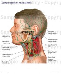
Jugular trunk on both sides joining the subclavian trunks. Both drain into the respective subclavian veins that both drain into the SVC.
|
|
|
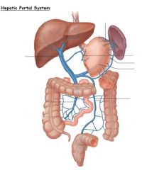
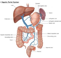
Going from inferior to superior, list the major veins draining the abdominal viscera into the portal vein
|
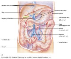
1) inferior mesenteric joining the spleni
2) superior mesenteric 3) gastro-epiploic anastomosing at spenic 4) gastric 5) cystic |
|
|
Starting at the celiac trunk moving inferiorly, describe the major abdominal arteries of the foregut
|
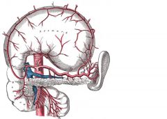
1) CT -> LG anastomosing at hepatic
2) CT -> splenic -> left gastro-epiploic anastomosing at right gastroepiploic 3) CT -> common hepatic -> gastroduodenal -> right gastro-epiploic 4) gastroduodenal -> superior pancreaticoduodenal |
|
|
What organs of the midgut are supplied by the superior mesenteric
|
1) 2nd part of duodenum down to first part transverse colon
2) head of pancreas |
|
|
List the organs of the hindgut supplied by the inferior mesenteric artery
|
2nd 2/3 of transverse colon to 1st 1/2 of rectum
|
|
|
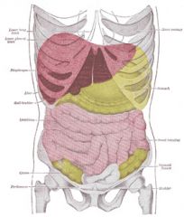
Describe the boundaries of the abdominal cavity
|
1) posterior abdominal wall
2) anterior & lateral: aponeurosis, transverse abdominis, rectus abdominis (divided by linea alba) and the aponeurosis, internal, external oblique |
|
|
Describe the origin, insertion, flexors and sheath of the rectus abdominis
|
Origin: pubic tubercles
Insertion: sternum and lower ribs Flexors: vertebral column Above arcuate line: rectus pair surrounded anterior and posterior by sheath made up equally of ex and int oblique and transverse abdominal Below arcuate line: all 3 muscles form anterior sheath of rectus pair, posterior is simply fascia. |
|
|
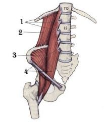
From posterior to anterior describe the muscles of the posterior abdominal wall
|
1) Quadratus lumboratum: 12th rib, lumbar transverse processes, iliac crest
2) psoas: origin t12 through sacrum and inserts at the femur |
|
|
describe the ligaments attached to the liver
|
1) bare area: coronary ligaments (anterior & posterior) joining right triangular. left triangular joining falciform
2) falciform separates left lobe from quadrate (inferior) and caudate (superior) lobes |
|
|
What are the main branches of the portal vein
|
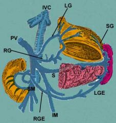
1) left gastric (lesser curvature)
2) splenic (left epiploic -> greater curvature) 3) superior and inferior mesenteric |
|
|
Describe the features of a hepatic lobule
|
1) Distally branches of the portal vein and artery drain into sinusoids which drain into a central vein that drains into the hepatic vein
2) Hepatocytes, straddling sinusoids and cannaliculi, excrete bile into cannaliculi and filter blood 3) Kupffer cells are macrophages within sinusoids |
|
|
Compare the anatomuy of the jejunum & ilium
|
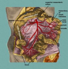
Jejunum: large distal arcades, long vasa recta, long numerous villi
Ilium: fatty mesentary, short vasa recta, fewer villi, lots of MALT |
|
|
describe the formation of "pouches" along the large intestine
|
Haustra formed by contraction of teniae coli.
|
|
|
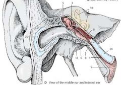
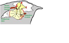
What are the main structures of the middle ear
|
1) Auditory ossicles: malleus, incus, stapes
2) Middle ear cavity 3) Tensor timpani (origin temporal bome, inserts malleus - CN5) 4) Stapedius (CN7) 5) Eustacian tube |
|
|
From the bottom up, describe the glands of the gastric gland
|
1) Chief cells
2) Endocrine glands (ECL, G) 3) Parietal cells 4) Mucous neck cells |
|
|
In between the lumen of the stomach and the muscularis externa, what are the main features
|
1) gastric pits
2) gastric epithelium (mucous secreting columnar) 3) Lamina propria 4) gastric glands (mucosal layer) 5) muscularis mucosa (submucosal plexus) 6) submucosa |
|
|
List the glandular cells prevalent in the pyloric antrum
|
1) ECL (histamine)
2) G cells (gastrin) 3) D cells (somatostatin) |
|
|
How is the tracheobroncheal tree supplied with blood
|
bronchial arteries directly from the aorta and from the intercostal arteries
|
|
|
How is the blood supplied and drained from the lungs
|
1) obtains oxygenated blood from the broncial artery and pulmonary vein (anatomic left to right shunt)
2) drains into azygous and hemiazygous veins |
|
Names
|
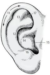
helix (A11), antihelix (A12), scapha (A13), concha (A14), tragus (A15), antitragus (A16), and triangular fossa (A17)
|
|
Names
|
mallear stria (B19) is caused by the attachment of the manubrium of the malleus, which reaches to the umbo of the tympanic membrane (B20), the innermost point of the funnel-shaped eardrum. Above the upper end of the mallear stria (mallear prominence) lies a lax, thin part of the eardrum, the reddish pars flaccida (B21), which is separated from the firm, gray and shiny pars tensa (B22) by two mallear folds.
|
|
Names
|
Auricle (D1), external acoustic meatus (D2), tympanic cavity
(D3), mastoid cells (air cells) (A6), otopharyngeal tube (D4) epitympanic recess (D5) above the external acoustic meatus, tympanic opening (D6). The tube extends obliquely downward and forward and opens in front of the posterior pharyngeal wall into the pharyngeal cavity (pharyngeal opening) (D7). isthmus of the tube (D8), tubal cartilage (D24), tensor tympani muscle (D9) attaches to the base of the manubrium |
|
Name them
|
saccule (AB6) and utricle (AB7). sensory epithelium
(blue), the macula of the saccule (AB8), macula of the utricle (AB9), interconnected by the utriculosaccular duct (AB10). endolymphatic duct (A11), the endolymphatic sac (A12). The uniting duct (AB13), scala media(AB15), the scala vestibuli (AB19), scala tympani (AB20), semicircular ducts (A22), common membranous crus (AB27) |
|
Name the parts
|
|
|
|
Describe the basic steps of lower respiratory embryological development
|
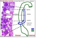
1) Laryngeotracheal tube derived from endoderm in pharynx, deepens to form lung bud
2) This splits into two bronchi at the carina 3) Mesenchyme accompanying the foregut derived mesenchyme forms the smooth muscle, nerves and blood vessels of the lungs. |
|
|
What is the difference between bronchi, terminal bronchioles, conducting broncioles and respiratory bronchioles
|
Bronchi: start of with C shaped rings of cartilage that become plates and have smooth muscle, surface is pseudostratified epithelium and glands
Conducting Bronchioles: smooth muscle, pseudostratified epithelium with goblets, no cartilage or glands Terminal bronchioles: smooth muscle, no glands, no goblet cells, ciliated cuboidal & clara cells (surfactant) Respiratory bronchioles: some smooth muscle, some ciliated cuboidal, more clara |
|
|
What are the 3 cells of the alveolus
|
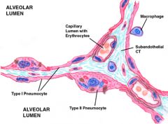
1) Type 1 pneumocyte (non-dividing respiratory, tight junctions)
2) Type 2: (dividing, matures into type 1, produces surfactant & lipid) 3) macrophages |
|
|
going by the order of receiving aortic blood flow first, name the 3 branches off the aorta
|
1) Cephalic trunk (-> branch right common carotid and subclavian)
2) Left common carotid 3) Left sub-clavian |
|
|
Describe the orientation of the aorta and pulmonary artery, allowing the ductus arteriosus to form
|
The aortic arch is directly over the pulmonary artery allowing an almost vertical shunt to form
|
|
|
What vessels enter the right atrium
|
Inferior & superior vena cavas
|
|
|
What vessels enter the left atrium
|
The 4 pulmonary veins
|
|
|
List the major coronary arteries
|
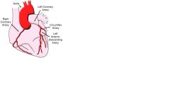
Left: LC (1cm) splits to L.circumflex and anterior descending atrioventricular
Right: RC branches into the circumflex |
|
|
Describe the origin of the coronary arteries
|
L & R coronary sinuses immediately beyond the aortic valve. The left one is identified by a larger osteum (entrance).
|
|
|
Describe how the coronary veins drain
|
Follow the anterior & posterior AV grooves to form the cononary sinus, draining into the RV below the tricuspid valve.
|
|
|
Find McBurney's point
|
Draw a line from the ASIS to the umbilicus. 1/3 way up.
|
|
|
What is the surface marking of the bifurcation of the abdominal aorta
|
Line joining both ASIS, midway
|
|
|
Define the transpyloric plane
|
Midway between the suprasternal notch & pubic synphisis. Level of L1
|
|
|
List 2 major blood vessels located at the transpyloric plane
|
superior mesenteric artery, renal arteries
|
|
|
List the organs indicated by the surface marking of the transpyloric plane
|
1) duodenojejunal flexure & 2nd part of duodenum, transverse colon
2) hylus' of the kidneys & pancreas 3) pylorus 4) neck of pancreas 5) fundus of gall bladder |
|
|
Describe the ductus venosus
|
Joins the umbilical vein to the IVC, bypassing the portal vein
|
|
|
List the 4 embryonic heart septation processes that occur
|
1) interventricular
2) atrial 3) atrioventricular 4) aorticopulminary (truncus arteriosus into aorta and pul trunk) |
|
|
Describe how the development of the atrial septum allows rapid changes to blood flow following birth
|
1) Septum primum and secundum develop with off-centred foramens
2) Septum primum on the left atrial side flaps open, allowing blood to pass through both foramens right to left pre birth 3) After birth, fall in pulmonary BP snaps the septum primum against the secundum, sealing the atrial septum. |
|
|
What are the 3 main anatomical abnormalities of tetralogy of Fallot
|
1) interventricular septum fails to complete
2) aorticopulmonary septum forms abnormally aorta causing it to overarch both ventricles 3) aorticopulmonary septum causes abnormally constricted pulmonary trunk - infundibular stenosis |
|
|
Describe the surface markings of the heart
|
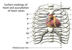
Lies between the 2nd rib (sternal angle) and the 5th intercostal space. The apex is pressed against the chest wall at the mid clavicular line.
|
|
|
Describe the outer coverings of the heart
|
1) Parietal pericardium - tough fibrous, fixed in several places
2) Visceral pericardium: inner epicardium joined to heart. 3) pericardial fliud between parietal & visceral pericardia |
|
|
Describe the main arteries of the right side of the heart
|
1) right coronary artery following the atrioventricular groove
2) marginal artery |
|
|
Describe the main arteries of the left side of the heart
|
1) left coronary artery
2) circumflex artery 3) anterior interventricular artery |
|
|
What are the main differences between the inflow and outflow valves of teh heart
|
Inflow: cuspid valves able to withstand maximal pressure of contraction via early contraction of chordae tendinae
Outflow: semilunar valves |
|
|
Describe the 2 types of sweat glands
|
1) apocrine: drain into hair follicles, produce cloudy odiferous sweat
2) merocrine: clear sweat directly mainly onto palms and soles of feet. |
|
|
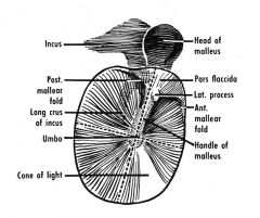
Describe the tympanic membrane
|

Membrane made up of three layers: stratified epithelium on the external side
cuboidal on the inner mucosa cartilage in the middle. At the anterior, inferior quadrant is the cone of lightadjacent to the central umbo of the malleus |
|
|
Describe (draw) the structures of the femoral triangle
|
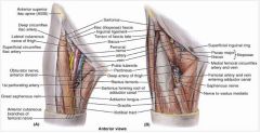
Know AVANS - adductor longus, vein, artery, nerve, sartoris
Posterior: pectineus (medial), iliacus (lateral) |
|
|
Describe the compartments of the mediastinum
|
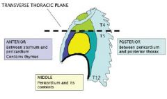
Superior (above T4/T5 = trans thoracic plane)
Inferior (below T4/T5) Anterior (thymus) Middle (pericardium) Posterior (azygous vein system |
|
|
Describe Waldeyer's ring
|
1) pharyngeal tonsil behind uvula adjacent to auditory tube
2) lingual tonsils at at base of tongue 3) palatine tonsils in oropharynx 4) |
|
|
Describe respiratory epithelium
|
1) ciliated pseudostratified columnar cells
2) goblet cells |
|
|
Describe the main parts of olfactory epithelium
|
1) olfactory epithelium
2) lamina propria containing Bowman's glands & nerve bundles 3) cribriform plate 4) olfactory bulb |
|
|
What is another name for the auditory tube and what cells line it
|
1) pharyngotympanic tube
2) respiratory epithelium: ciliated pseudostratified + goblet cells |
|
|
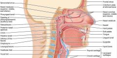
Draw the nasal, oral cavities, pharynx, larynx, oesophagus & trachea. Mark the cervical vertebral levels
|
Label: nasal vestibule, olfactory epithelium (OE, lamina propria & Bowman's glands, crib plate, olfac bulb), opening to auditory tube, oropharynx from C4 up, epiglottis, larynx & laryngopharynx from C6 to c4, trachea & oesophagus
|
|
|
Name the main parts of the pinna
|
helix, concha, tragus & antitragus, EAM, lobule
|
|
|
Name the 2 muscles involved in the acoustic reflex
|
1) tensor tympani
2) stapedius |
|
|
What are the layers of the tympanic membrane and the epithelium of the middle ear
|
1) stratified epithelium, fibrocartilage, simple cuboidal
2) inner ear is simple cuboidal |
|
|
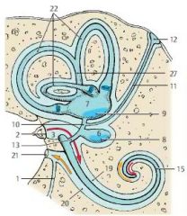
Name the main parts of the inner ear
|
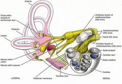
1) cochlea (contains organ of Corti, scala media, scala tympani, scala vestibuli, tectorial membrane) - cochlear nerve
2) semicircular canals and ducts 3) vestibiule: utricule and saccule for gravity and inertia - has 2 vestibular nerves (vestibulocochlear nerve CN VIII) |
|
|
You have to perform a needle thoracentesis. What tissues does the needle have to penetrate & where should the needle be placed
|
1) skin -> fascia -> serratus anterior -> external intercostal -> internal intercostal -> innermost intercostal -> parietal pleura
2) immediately above the lowe rib to avoid damage to the intercostal artery, vein & nerve |
|
|
What muscles are used for forceful expiration
|
1) internal intercostals
2) abdominal: external oblique, internal oblique, transversus abdominis |
|
Name the bits
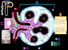
|
A – Renal corpuscle
B – Proximal tubule C – Distal convoluted tubule D – Juxtaglomerular apparatus 1. Basement membrane (Basal lamina) 2. Bowman's capsule – parietal layer 3. Bowman's capsule – visceral layer 3a. Pedicels (Foot processes from podocytes) 3b. Podocyte 4. Bowman's space (urinary space) 5a. Mesangium – Intraglomerular cell 5b. Mesangium – Extraglomerular cell 6. Granular cells (Juxtaglomerular cells) 7. Macula densa 8. Myocytes (smooth muscle) 9. Afferent arteriole 10. Glomerulus Capillaries 11. Efferent arteriole |
|
|
What are the divisions on an ECG
|
5mm = 0.5mv
5mm = 0.2s |
|
|
Describe the P wave of an ECG
|
1) atrial depolarisation right to left (+ve in leads I-III, AVL, AVF, -ve in AVR)
2) about 100ms (2.5 divs) |
|
|
Describe the Q wave of an ECG
|
septal depolarisation from left to right (-ve in leads I-III, AVL, AVF, +ve in AVR)
|
|
|
Describe the R wave of an ECG
|
1) left ventricular depolarisation right to left (+ve in leads I-III, AVL, AVF, -ve in AVR)
2) about 100ms (2.5 divs) |
|
|
Describe the S wave on an ECG
|
septal repolarisation from right to left (-ve in leads I-III, AVL, AVF, +ve in AVR)
|
|
|
Describe the structures of the superior mediastinum
|
1) great vessels: superior VC dividing into the L&R brachiocephalic veins, pulmonary trunk & veins
2) ABCs from right to left: aorta, brachiocephalic artery, left common carotid, left subclavian 3) phrenic & vagus nerves 4) trachea (bifurcation at T4/T5 = angle of Louis, also thymus) & oesophagus |
|
|
Describe the structures of the anterior, middle & posterior mediastinum
|
anterior: thymus
middle: heart posterior: oesopahgus, vagus nerve branches, azygous vein |
|
|
to reside
|
abitare
|
|
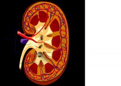
Label the parts of the kidney
|
1.Renal pyramid
2.Interlobar artery 3.Renal artery 4.Renal vein 5.Renal hilum 6.Renal pelvis 7.Ureter 8.Minor calyx 9.Renal capsule 10.Inferior renal capsule 11.Superior renal capsule 12.Interlobar vein 13.Nephron 14.Minor calyx 15.Major calyx 16.Renal papilla 17.Renal column |
|
|
Explain why a large volume of urine is excreted in the recovery phase of ATN
|
Tubules allowing passage of filtrate but cannot reabsorb sufficient water ie: 180L/day of filtrate must be reabsorbed: 70% in proximal, 20% in loop, 9% in distal tubule & collecting duct
|
|
|
How is sodium reabsorbed in the proximal tubule
|
Secondary active transport of: 1) the Na+/H+ antiporter reabsorbs both bicarbonate and sodium
2) SGLT-1: sodium/glucose symporter 3) solvent drag |
|
|
What is the main difference between the function of the proximal convoluted & straight tubule
|
Proximal convoluted: reabsorption
Proximal straight: secretion of weak acids (eg B-lactam antibiotics) |
|
|
How does the thick ascending limb of the LOH dilute urine
|
Secondary active transport of NAKCl2 symporter dragging H2O. K+ is allowed into interstitium to maintain hyperosmolarity
|
|
|
Where is Ca++ reabsorbed in the nephron and how is this regulated
|
Distal convoluted tubule. Reabsorption increased by PTH
|
|
|
What is the principle ion transport mechanism of the distal tubule
|
1) Na+/K+ active transport on basolateral membrane drives Na+/Cl- symporter on apical membrane (thiazides bind here)
2) Na+/Ca++ active transport on basolateral membrane drives Ca+ secondary transport. |
|
|
What is the principle ion transport mechanism of the late distal/collecting ducts
|
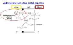
ENaC Na channel driven by Na+K+ ATPase. Activated by aldosterone, blocked by amiloride
|
|
|
Which cells of the collecting duct produce HCO3- de-novo
|
Intercalated cells secreting H+
|
|
|
Describe the physiology of bladder filling & micturition
|
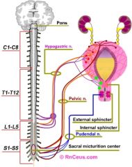
1) hypogastric nerve (T12/L1) counters PNS tone of bladder detruser (B-receptors) & contracts internal sphincter (a-receptors). Pudental nerve constricts external sphincter
2) Stretch of detruser by >150mL increases pelvic nerve tone: detruser contraction. Pudental & hypogastric nerves inhibited supraspinally, opening both sphincters. |
|
|
List the features of the right atrium
|
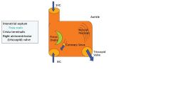
1) sup & inf vena cavas
2) fossa ovale (SA node) 3) coronary sinys 4) pectinate muscles |
|
|
List the features of the right ventricle
|
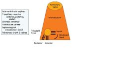
1) pulmonary semilunar valve
2) tricuspid valve with 3 chordae tendinae & papillary muscles |
|
|
Liist the features of the left atrium
|
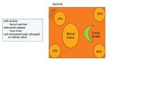
1) openings from the 4 pulmonary veins
2) fossa ovale 3) superior face of the mitral valve |
|
|
List the features of the left ventricle
|
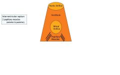
1) aortic valve
2) vestibule 3) mitral valve, 2 chordae tendinae & papillary muscles |
|
|
Describe the omenta of the stomach
|
1) greater (loops to transverse colon) & lesser omenta
2) gastrosplenic 3) hepatoduodenal (extension of lesser omentum - only to 1st part & contains portal triad) 4) gastrophrenic (joins fundus to diaphragm) |
|
|
What are the maiin posterior relations of the stomach
|
lesser sac
transverse mesocolon spleen pancreas left kidney |
|
|
Describe the vertebral levels of the duodenum
|
1st part, up from L1 and lateral right
2nd part: vertical down L1-L3 3rd part: across left L3 and up 4th part: L2 and anterior to join jejunum which is intraperitoneal |
|
|
Describe the layers of the stomach
|
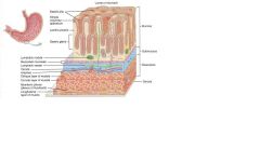
mucosa( mucous epithelium, lammina propria, muscularis mucosa)
submucosa muscularis externa (oblique, circular, myenteric plexus, longtitudinal) |
|
|
Describe the 5 types & locations of specialised cells of the stomach
|
1) mucous
2) mucous neck cells 3) chief cells (base of gastric pit- pepsinogen) 4) parietal cells (neck of gastric pit) 5) endocrine cells: G=gastrin, ECL=histamine, D (somatostatin) |
|
|
List 3 types of obstruction of tubes with examples
|
1) extra tubular (pressure from tumours, fibrosis)
2) tubular (stricture, tumour, neuromuscular (absent peristalsis) 3) luminal (secretions eg CF, stones eg renal calculi, cholelythiasis, thrombi & emboli |
|
|
Describe the gross anatomy of the spleen, including blood vessels
|

1) head (unicate process), neck body, tail
2) pancreatic duct drains into superior 2nd part of duodenum via the ampulla of Vater 3) body and tail supplied by splenic artery/vein 4) head supplied by pancreaticoduodenal arteries |