![]()
![]()
![]()
Use LEFT and RIGHT arrow keys to navigate between flashcards;
Use UP and DOWN arrow keys to flip the card;
H to show hint;
A reads text to speech;
74 Cards in this Set
- Front
- Back
|
Describe the structure of the pericardium. What structures does it cover? |
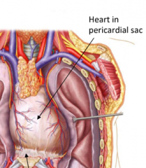
The Pericardium is a fibroserous membrane. The pericardium covers the heart and the great vessel roots |
|
|
What are the two layers of the pericardium and how are they arranged? |
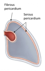
Two layers of pericardium: - Fibrous - Serous |
|
|
What are two 'layers' does the serous pericardium form? |
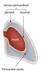
The serous pericardium comprises the parietal (outer) and visceral (inner) layers as one continous serous membrane. The cavity formed is the pericardial cavity (potential space), containing small amount of fluid to reduce friction. |
|
|
The apex of the heart lies on which space of the heart? Which chamber forms the apex? |
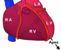
The apex of the heart is located on the left 5th intercostal space. It is formed by the left ventricle |
|
|
Where is the base of the heart located? |
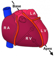
On the posterior aspect of the heart. |
|
Each Atrium forms an "ear shaped" appendage/structure which projects to teh front of the heart. What is this called? |

The Auricles |
|
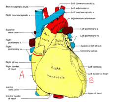
Label structures A and B on the anterior surface of the heart. |
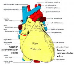
A= Anterior atrioventricular sulcus (both continue onto posterior side of the heart) |
|
|
The VC feeds into the Right Atrium. Which areas of the body does the superior and inferior VC receive venous return from? |
Superior VC: - All drainage from above, (thorax, head, etc) Inferior VC: - All drainage from below (abdomen, pelvis, lower limbs etc.) |
|
|
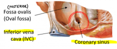
What is the function of the coronary sinus, and where is it located in the heart? |
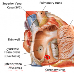
The coronary sinus receives all venous return from the heart itself. It drains into the Right Atrium and it's located beside the IVC on the lower rear side. |
|
|
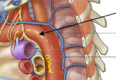
What is the Fossa Ovalis (oval fossa) and what is it remnant of? |
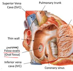
The fossa ovalis is a thin fibrous sheet between the two atria. It is remnant of the foramen ovale which allowed blood to pass from RA --> LA in fetal development |
|
|
Which valve lies between the RIGHT ATRIUM and the RIGHT VENTRICLE? What structures attach to this valve? |
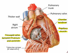
The Atrioventricular tricuspid valve The chordae tendinae and papillary muscles |
|
|
Papillary muscles and chordae tendinae only attach to which types of valves? How do these structures assist the prevention of backflow and what direction do they contract towards? |
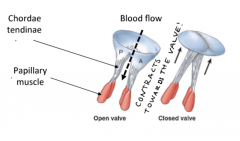
only attach to atrioventricular valves. Prevent backflow by forced closure due to pressure gradient in the ventricular contraction. They contract towards the valve. |
|
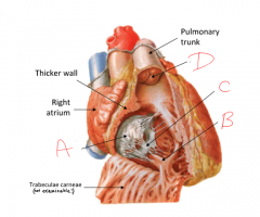
Label the structures A-D shown |
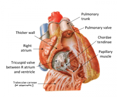
A= Tricuspid Valve B= Papillary muscle C= Chordae tendinae D= Pulmonary valve |
|
|
The left ventricle is separated from the left atrium by which valve? In terms of the wall thickness, describe the left ventricular walls? |
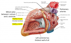
Mitral/bicuspid Valve. |
|
|
The left atrium Contains an oval shaped impressino on its inner wall (adjacent to the right atrium) - what is the name of this structure? |
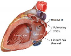
Fossa ovalis |
|
|
The fibrous skeleton of the heart is comprised of what two paired components? |
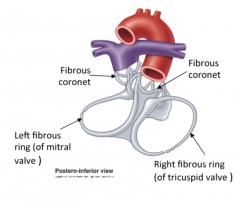
Composed of two fibrous rings and two fibrous coronets. Together they provide: - attachment for myocardium - a form of electrical insulation --> separating the atrial muscles from the ventricular muscle |
|
|
The pulmonary and aortic valve lie where? What is there structure often referred to as? |
Pulmonary lies at the beginning of the pulmonary artery in the right ventricle (exit) The aortic valve lies at the beginning of the aorta in the left ventricle (exit) They are often referred to as 'semilunar' valves The closure and opening of these is passive |
|
|
Where are the orifices of the coronary arteries found? |
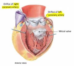
Superior to the aortic valve. |
|
|
What is the sinoatrial (SA) node commonly called? |
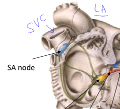
The SA node is located at the junction of the: superior vena cava and the right atrium. It is often called the pacemaker of the heart. |
|
|
Where would you rind the atrioventricular (AV) node? What is its function? |
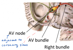
in the r-atrium, near the opening of the coronary sinuses and close to the cusps of the tricuspid valves.
the AV node distributes electrical signals to ventricles via AV bundles |
|
|
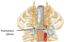
How is the heart innervated by nerve? |
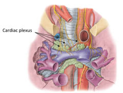
The heart is innervated by autonomic fibres from cardiac plexus (base of heart). Both parasymp and symp fibres. |
|
|
Where does the ascending aorta arise from? |
The left ventricle |
|
|
Where is the aortic arch located in relation to the body?. Describe the direction the vessel initially travels. |
Located at level of manubrial-sternal junction. Arches superiorly, posteriorly and to the left. |
|
|
Does the aortic arch travel over or under the bifurication of the trachea? |
Travels over the trachea. Leaves impression on left lung |
|
|
At what vertebral level (roughly) does the descending aorta align medially? |
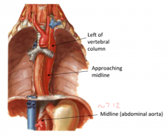
roughly at T12 (abdominal aorta) |
|
|
Where are the coronary arteries found? |
Located: superior to aortic valve RCA: supplies R-atria + R-ventricle LCA: supplies L-atria + L-ventricle |
|

What is branch is structure 'A' of the aortic arch? |
Brachiocephalic trunk (branches into subclavian + common carotid) |
|

What is branch is structure 'B' of the aortic arch? |
Right subclavian artery |
|

What is branch is structure 'C' of the aortic arch? |
Right carotid artery |
|

What is branch is structure 'D' of the aortic arch? |
Left common carotid artery |
|
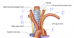
What is branch is structure 'E' of the aortic arch? |
Left subclavian artery |
|
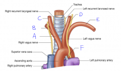
What is structure 'F'? what is it remnant of? |
Ligamentum arteriosum. Remnant of the ductus arteriosis in fetal circulation |
|
|
What are some areas supplied by the parietal branches of the descending aorta? |
Parietal: - intercostal = walls - pericardial = pericardium - phrenic = diaphram |
|
|
What are some areas supplied by the visceral branches of the descending aorta? |
Visceral: - Bronchial = airways, bronchial trees - Esophageal = esophagus |
|

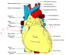
In the image depicting the 'great veins' what are structures 'A'? What feature regarding its position affects the venous return rate? |

A = brachiocephalic veins It slightly to the right meaning that the right side drains slightly faster (less distance to travel) |
|

In the image depicting the 'great veins' what are structures 'B'? which arteries to the travel alongside and where do they return venous flow from? |
B = internal jugular veins accompany the common carotid arteries return venous flow from the head and neck |
|
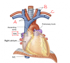
In the image depicting the 'great veins' what is structures 'C'? (should be labelled for both sides) |

C= subclavian veins accompanies the subclavian arteries |
|
|
Do the coronary veins accompany the coronary arteries? What structure do they drain into and where is this located? |
Yes the coronary veins accompany the coronary arteries. They drain into the coronary sinus which is located next to the IVC |
|
|
Where does the azygous vein receive it's blood return from? What structure does it arch over? |
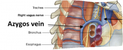
The azygous vein receives all venous return from : - thoracic wall and pericardium - lungs, trachea, bronchi and esophagus. it arches over the root of the right lung |
|
|
Which lung does the azygous vein leave an impresion on? (azygous groove?) |
the right lung |
|
|
What artery supplies the AV and SA node? are they functional end arteries?
|
Coronary arteries. Yes the coronary arteries are functional end arteries |
|
|
Where do the right coronary arteries lie? (assume classic distribution) |
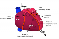
Lies in the atrio-ventricular groove. Follows groove onto posterior side where it anastamoses with the left coronary artery. |
|
|
What two branches does the left coronary artery branch into? where do each of these structures lie on the heart and where do they anastamose? |

Divides into: Circumflex branch: |
|
|
If you were to suddenly block part of the coronary artery, would the anastamoses be able to compensate for obstruciton of flow? |
Sudden block = not be able to compensate --> death |
|
|
The thoracic cavity is divided in to 3 major compartments which are? |
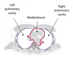
- Right pulmonary cavity (right lung site) - Medianastium (compartment between two lungs) - left pulmonary cavity (left lung site). |
|
|
Describe the structure of the pleura |
Pleura = continuous serous membrane that covers each lung and the wall of the pleural cavity. |
|
|
What 2 sections can the pleura be divided into and what structures do they cover? |
Can be divided into: Parietal pleura: covers wall of pleural cavity visceral pleura: covers surface of each lung |
|
|
What four sections can the parietal pleura be divided into? What do they relate to/surround? |
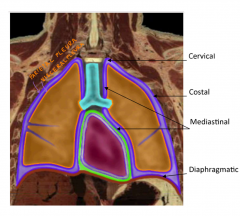
1. cervical pleura - lines cervical extension of pleural cavity 2. costal pleura - related to ribs and intercostal space 3. mediastinal pleura - covers mediastinum 4. diaphragmatic pleura - covers the diaphragm |
|
|
What is the purpose of the pleural cavity? Does it contain fluid? |
Purpose: creates frictionless environment Fluid: yes, few mls of serous fluid It is only a potential space between visceral and pleural cavity |
|
|
What are the two key characteristics of the pulmonary ligament, and what structure does it form? Does it lie superior or inferior to the lung root? |
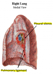
1. Double-folded pleura 2. Represents a continuity between the parietal and visceral pleura - Forms the pleural sleeve - Lies inferior to the lung root |
|
|
What type of nerve innervation do each of the visceral and parietal pleurae receive? |
Visceral pleura receive autonomic nerve innervation Parietal pleura receive somatic nerve innervation. |
|
|
What is the clinical significance of penetration of the pleural cavity? |
The pleural cavity is supplied by somatic nerves, and hence is extremely painful during needle-penetration procedures. |
|
|
What two sections enclose the trachea and what are they comprised of?
|
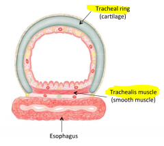
Tracheal ring (cartilage) - horseshoe shape Trachealis muscles (smooth muscle) - encloses posterior surface |
|
|
At what vertebral level does the bronchial tree begin and at what level does the bronchus divide into the right and left bronchus? |
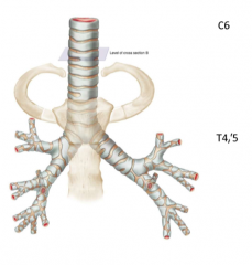
C6 |
|
|
When comparing the right main bronchus to the left main bronchus, which is larger and more medial? Consequently, which is more prone to obstruction? |
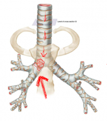
The right main bronchus is: - Wider and hence is more likely to be obstructed than the left main bronchus |
|
|
What do the main bronchus subdivide into? what does this further subdivide into? |
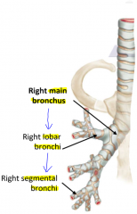
Main bronchus --> lobar bronchi --> segmental bronchi |
|
|
What are the 3 characteristics of the individual bronchopulmnoary segments? |
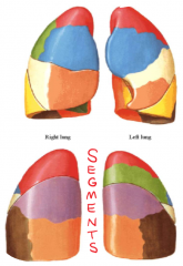
1. Each supplied by independently segmented bronchus 2. Each have independent blood supply 3. Surgically resectable |
|

What is structure '1' of the right lung? (anterior view) |

Apex of the lung |
|

What is structure '2' of the right lung? (anterior view) |

Superior lobe |
|

What is structure '3' of the right lung? (anterior view) |
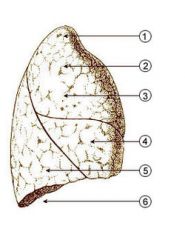
Costal surface |
|

What is structure '4' of the right lung? (anterior view) |

Middle lobe |
|

What is structure '5' of the right lung? (anterior view) |

Inferior Lobe |
|

What is structure '6' of the right lung? (anterior view) |

Base or diaphragmatic surface |
|

What is structure 1 of the right lung? (medial view) |

Anterior border |
|

What is structure 2 of the right lung? (medial view) |
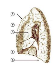
Mediastinal part (could also be groove for azygous vein) |
|

What is surface 3 of the right lung? (medial view) |

Medial surface |
|
|
What are the 4 key difference between the left and right lung? 1. fissures? 3. main bronchi? 4. size/length? |
1. Left lung only has oblique fissure (superior and inferior lobes) 2. Groove on left lung is due to aortic arch Groove on right lung due to azygous vein 3. Right main brochus has already started to divide at the hilum, whereas the left main bronchus has not yet split. 4. right lung is bigger and heavier, however it is shorter. The left lung also has the cardiac notch, (where the heart sits. |
|
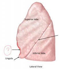
On the left lung shown below, what is labelled? what forms this indent? |
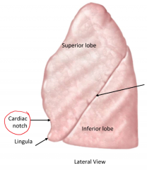
Cardiac notch. (impression left by heart.) |
|
|
Which arteries and veins are responsible for oxygenation in the lungs? |
Pulmonary arteries and veins |
|
|
Which arteries and veins are responsible for vascular supply of the lungs? |
The bronchial arteries and veins |
|
|
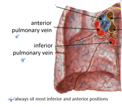
Within the structures of the hilum of each lung, what positions do the pulmonary arteries lie? |
In the inferior and anterior most positions |
|
|
When comparing the position of the pulmonary vein in both the right and left lung what is the key difference? |
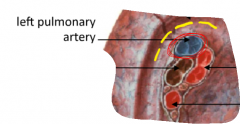
in the Left lung, the pulmonary artery always sits in the most superior position. (right lung is more central and lower. |
|
|
Describe the 4 stages of lymph drainage (pathway) of the lungs |
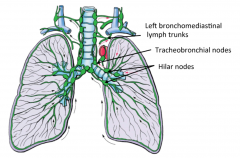
1. superficial and deep lymphatics 2. Bronchopulmonary (hilar nodes) 3. Tracheobronchial nodes 4. Bronchomediastinal lymph trunks (right or left) |
|
|
Where do the lungs and visceral pleurae receive nerve innervation from? |
Lungs and visceral pleurae receive nerve innervation from the pulmonary plexus Supplied by both parasymp. and symp. |