Reading...
![]()
Play button
![]()
Play button
![]()
Use LEFT and RIGHT arrow keys to navigate between flashcards;
Use UP and DOWN arrow keys to flip the card;
H to show hint;
A reads text to speech;
166 Cards in this Set
- Front
- Back
|
1 a. What is the relationship between a joint’s mobility and stability?
|
The more mobile a joint the less stable it is.
|
|
|
b. What is the least stable joint? Why? What is the most mobile joint?
|
The shoulder is the most unstable joint. It has the widest range of motion making it the most mobile joint as well
|
|
|
2 a. Describe the structural differences between fibrous and cartilaginous joints.
|
fibrous joints have dense fibroustissue. Cartilaginous jointsare joined by Cartilage
|
|
|
b. Describe the functional classification of the fibrous and cartilaginous joints.
|
Fibrous jointsare immovable or slightly movable. I.e.the joints of the skull. Cartilaginous joint are also notable to move but are flexible.
|
|
|
3 a. What is synarthrosis? Give some examples.
|
Synarthrosis is an immovable joint.Sutures of the skull.
|
|
|
b. Why do we need synarthrosis?
|
the joint need flexibility but stability as well
|
|
|
4 a. Discuss the origin and function of synovial fluid.
|
the synovial joint joins two long bones and provides a fluid inside the synovial memebrane for lubrication during movement.
|
|
|
b. Why is there no synovial fluid in other types of joints?
|
because lubrication for movement is not required in those joints.
|
|
|
5 a. Compare a hinge joint and a pivot joint with respect to structure, function, and location.
|
a hinge joint allow for angular movement along a single plane (i.e. Knee or elbow). A Pivot joint permits rotation along an axis (the joint between the atlas and axis)
|
|
|
b. Describe one similarity between the upper and lower limbs with respect to joints?
|
Both the lower and upper limbs have hinge joints that allow movement for locomotion
|
|
|
6 a. Compare a saddle joint and a condylar joint with respect to structure, function, and location.
|
A saddle join resembles a saddle with one concave end and cone convex end. Saddle joints allow for angular motion without rotation. The base of the thumb is an example of of a saddle joint . A condylar joint , also know as an ellipsoidal joint, has an oval face that nestles within a depression on the opposing surface. Condylar joints allow for angular motion across two planes. They are an example of a biaxial joint and are found in the joints that connect the fingers and the toes with the metacarpal bones.
|
|
|
7 a. Describe the different types of functional classification of joints.
|
Joint are classified in 3 ways (1) the range of motion that they provide (2) the type of connective tissue between them (3) where the space between the occurs.
|
|
|
b. List one advantage to the body to have these different types of joints.
|
Having these 3 types of joints allows for a large range of movements and functions in the joints.
|
|
|
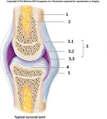
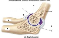
8 a. Describe the basic anatomy of a synovial joint.
|
(1) joint capusle (2) articualr cartilage (3) a joint cavity (4) a synovial (5) accesory structures (6) sensory nerves and blood vessels
|
|
|
b. Why is synovial joint the most abundant type in the body?
|
It is the joint that is found in any part of the body that moves.
|
|
|
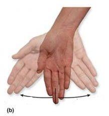
9 a. Describe and compare the movement of abduction to adduction and pronation to supination.
|
Abduction is the movement away from the longitudinal axis of the body. Adduction is movement of toward the longitudinal axis of the body. Pronation is the rotaion of a forward facing surface (with respect to anatomical position) backwards. Supination is the roation of a posterior facing surface forward,
|
|
|
b. Why is it advantagious to describe movements in opposing action?
|
It allow for a comparion of the movements base i=on their range of motion
|
|
|
10 a. Describe the anatomy of the intervertebral joints.
|
Invertebral joint are found in between the bones of the vertibral column.
|
|
|
b. Why do we need so many ligaments?
|
to hold the vertbrae in place because the joints are inherently unstable.
|
|
|
Articulation
|
Exist where ever two bones meet
|
|
|
Fibrous
|
"A fibrous joint occurs where bones are held together by dense regular (fibrous) connective tissue.
|
|
|
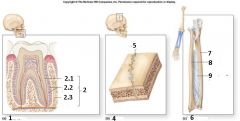
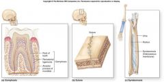
Gomphosis
|
a specialized form of fibrous synarthosis that binds each tooth to the surrounding bony socket. Eg periodonal ligaments between the teeth and jaws.
|
|
|
Periodontal Membrane
|
the fibourous connection in gomphosis
|
|
|
Sutures
|
a synarthrotic joint oly found in the skull -eg. The bones of the skull
|
|
|
Synostoses
|
A totally rigid immovable joint, where two bones fuse together and the boundry between them disappears
|
|
|
Syndesmoses
|
the articulaing bones are connected by a ligament that limits movement of the articulating bones.
|
|
|
Interosseous Membrane
|
is a broad and thin plane of fibrous tissue that separates many of the bones of the body. It is an important component of many joints.
|
|
|
Cartilaginous
|
cartilaginous joint occurs where bones are joined by cartilage.
|
|
|
Synchondroses
|
type of synarthroses when a diaphysis and epiphysis are bound together by an epiphyseal cartilage - eg. Epiphyseal cartilages
|
|
|
Epiphyseal Plate
|
a hyaline cartilage plate in the metaphysis at each end of a long bone. The plate is found in children and adolescents; in adults, who have stopped growing, the plate is replaced by an epiphyseal line.
|
|
|
Costochondral Joints
|
the articulations between the ribs and costal cartilage. They are hyaline cartilagenous joints
|
|
|
Symphysis
|
is a fibrocartilaginous fusion between two bones. It is a type of cartilaginous joint, specifically a secondary cartilaginous joint. i.e. pubic symphysis
|
|
|
Pubic Symphysis
|
is the midline cartilaginous joint (secondary cartilaginous) uniting the superior rami of the left and right pubic bones. It is located anterior to the urinary bladder and superior to the external genitalia; for females it is above the vulva and for males it is above the penis.
|
|
|
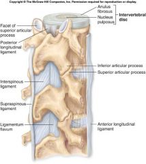
Intervertebral Disc
|
lie between adjacent vertebrae in the spine. Each disc forms a cartilaginous joint to allow slight movement of the vertebrae, and acts as a ligament to hold the vertebrae together. Discs consist of an outer annulus fibrosus, which surrounds the inner nucleus pulposus. the annulus fibrosus consists of several layers of fibrocartilage
|
|
|
Synovial
|
has a fluid-filled synovial cavity, bones are enclosed within a capsule, bones are joined by various ligaments
|
|
|
Uniaxial
|
the one only moves in one plane.
|
|
|
Plane Joint
|
gliding joints i.e.
|
|
|
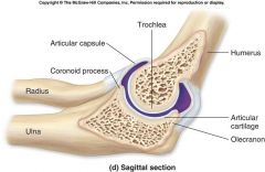
Hinge Joint
|
a bone joint in which the articular surfaces are moulded to each other in such a manner as to permit motion only in one plane—backward and forward. i.e. knee, elbow
|
|
|
Pivot Joint
|
a joint that moves by rotating. i.e. the axis/atlas, .
|
|
|
Biaxial
|
a bone that move in two planes
|
|
|
Condylar Joint
|
one in which an ovoid head of one bone moves in an elliptical cavity of another, permitting all movements except axial rotation.
|
|
|
Saddle Joint
|
Saddle joints, which resemble a saddle, permit the same movements as the condyloid joints.
|
|
|
Multiaxial
|
|
|
|
Ball-n-Socket Joint
|
These allow a wide range of movement.
|
|
|
Synarthrosis (Functional Classification)
|
an immovable joint- kinds fibrous, cartilaginous, and bony fusion.
|
|
|
Amphiarthrosis (Functional Classification)
|
a slightly movable joint - kinds Fibrous and cartilaginous
|
|
|
Diarthrosis (Functional Classification)
|
a freely moveable joint that permits a wide range of movement - also know as synovial joints
|
|
|
Synovial Joints
|
also know as diarthrosis, are spcialized for movementand permit a wide range of motion. I.e the shoulder joint, knee joint and the TMJ
|
|
|
Articular/Joint Capsule
|
thick layer of dense connective tissue that surrounds a synovial joint.
|
|
|
what structures are found in the typical synovial joint
|
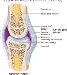
(1) joint capusle (2) articualr cartilage (3) a joint cavity (4) a synovial (5) accesory structures (6) sensory nerves and blood vessels
|
|
|
Synovial Membrane
|
line a joint but stops at the edges of the articular cavity. They produce synovial fluid.
|
|
|
Articular/Joint Cavity
|
capsule + synovial membrane. The fibrous capsule is continuous with the periosteum of bone. It is also highly innervated but avascular (lacking blood and lymph vessels)
|
|
|
Synovial Fluid
|
a fluid produced by the synovial membrane that servers 3 functions (1) lubrication (2) Nourshment of chondrocytes (3) shock absorber
|
|
|
Articular Cartilage
|
cartilage found in between bony surfaces of a synovial joint. Typically found at the end of long bones.
|
|
|
Ligaments
|
joint capsule that surrounds the entire joint and is continous with the periostea of the articualting bone.
|
|
|
Extrinsic ligaments
|
located inside or outside the joint capsule and are separate from the joint capsule
|
|
|
Intrinsic ligaments
|
a localized thickening of the joint capsule AKA capsular ligaments
|
|
|
Tendons
|
Connects muscle to bone, usually not part of the articulation but pass across or around a joint.
|
|
|
Bursa
|
small, fluid filled pockets of connective tissue.
|
|
|
Tendon Sheath
|
tubular bursea that surround tendons when they pass across bony surfaces.
|
|
|
Fat Pads
|
Found around the periphery of the joint , lightly covered by a layer of synovial memebrane. Provides protection for articular cartilages.
|
|
|
Gliding
|
also known as plane joints have flattened or slightly curved faces. Can be mutliaxial or nonaxial.
|
|
|
Angular Motion
|
movement in reference to 2 axis ie forward, backard and left and right
|
|
|
Flexion
|
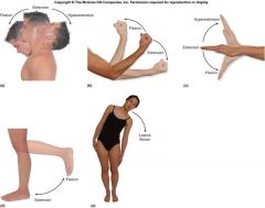
Flexion decreases the angle between the bones of the limb at a joint, while extension increases it.
|
|
|
Lateral Flexion
|
when the veterbral column bends to the side
|
|
|
Extension
|
straightening limbs at a joint.
|
|
|
Hyperextension
|
when part of the body is bent and overstretched or bent beyond it's normal range.
|
|
|
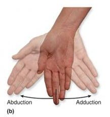
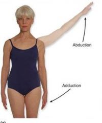
Abduction
|

movement away from the mid-line of the body.
|
|
|
Adduction
|
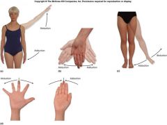
movement towards the mid-line of the body.
|
|
|
Circumduction
|
any type of angular motion
|
|
|
Rotational Motion
|
|
|
|
Rotation
|
a circular movement around a fixed point.
|
|
|
Lateral
|
is rotation away from the center of the body
|
|
|
Medial
|
is rotation towards the center of the body
|
|
|
Pronation
|
The movement of a limb away from the body
|
|
|
Supination
|
the return of the limb towards the body
|
|
|
Depression
|
occurs when a structure moves in an inferior direction. An example, would be when you close your mandible
|
|
|
Elevation
|
occurs when a structure moves in a superior direction. An example would be when opening the mouth the mandible is depressed.
|
|
|
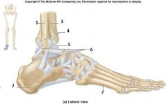
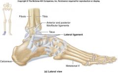
Dorsiflexion
|
ankle flexion elevates the distal of the portion of the foot and the toes. An example would be digging the heels in the foot.
|
|
|
Plantar Flexion
|
refers to movement of the foot. For example, standing on tip toe.
|
|
|
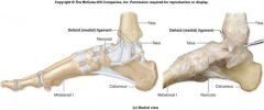
Inversion
|
is a twisting motion of the foot that turns the sole inward.
|
|
|
Eversion
|
is a twisting motion of the foot that turns the sole outward.
|
|
|
Protraction
|
entails moving a part of the body anteriorly in the horizontal plane. You protract your jaw when you grasp your upper lip with your lower teeth
|
|
|
Retraction
|
reverse movement of protraction. You protract your clavicles when you cross your arms.
|
|
|
Opposition
|
is the special movement of the thumb that produces pad-to-pad contact of the thumb with the palm or any other finger.
|
|
|
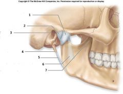
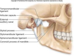
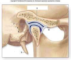
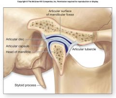
Temporomandibular Joint (TMJ)
|
is a small but complex multiaxial articulation between the mandibular fossa of the temporal bone and the condylar process of the mandible
|
|
|
Articular Capsule
|
surrounds the joint complex and isn’t well defined. The portion superior to the neck of the condyle is loose while the portion of the capsule is inferior to the cartilage disc is tight.
|
|
|
Articular Disc
|
pads of fibrous cartilage that may sudivide a synovial cavity. (1) channels the flow of synovial fluid (2) allows for variations of shape (3) or restricts movement at the joint.
|
|
|
Sphenomandibular Ligament
|

is a flat, thin band which is attached above to the spina angularis of the sphenoid bone, and, becoming broader as it descends, is fixed to the lingula of the mandibular foramen. The function of the sphenomandibular ligament is to limit distension of the mandible in an inferior direction.
|
|
|
Stylomandibular Ligament
|

is a specialized band of the cervical fascia, which extends from near the apex of the styloid process of the temporal bone to the angle and posterior border of the angle of the mandible, between the Masseter and Pterygoideus internus.
|
|
|
Temporomandibular/Lateral Ligament
|

is a small complex multilateral articulations between the mandibular fossa of the temporal bone and the condyler process of the mandible.
|
|
|
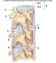
Intervertebral Articulations
|
located between the superior and inferior articular processs of the adjacent vertebras are plane joints that permit shall movements associated with flexion and extension.
|
|
|
Anulus Fibrosus
|
the tough outer layer of fibrous cartilage which surrounds the second part of the interebral disc.
|
|
|
Nucleus Pulposus
|
is the second part of the intervertebral disc which is composed of a soft, elastic gelatinous core, composed primarily of water.
|
|
|
Anterior Longitudinal Ligament
|
connects to the anterior surfaces of each vertebral body.
|
|
|
Posterior Longitudinal Ligament
|
parallels with the anterior longitudinal ligament but passes across the posterior surfaces of each body.
|
|
|
Interspinous Ligaments
|
the spinous processes of adjacent vertebrae.
|
|
|
Supraspinous Ligaments
|
interconnects the tips of the spinous process from C7 to the sacrum.
|
|
|
Ligamentum Nuchae
|
is a supraspinous ligament that extends from c7 to the base of the skull.
|
|
|
Ligamentum Flavum
|
is a ring of dense irregular CT that is attached to the joint cavity by fibrous cartilage.
|
|
|
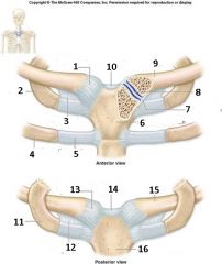
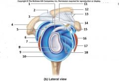
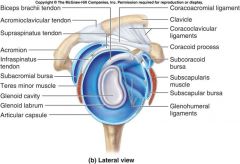
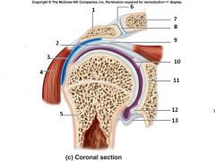
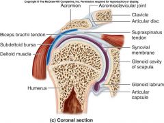
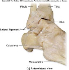
Glenohumeral (Shoulder) Joint
|
is a loose and shallow joint that permits the greatest range of motion of any joint in the body.
|
|
|
Glenoid Labrum
|
lip / edge which deepens the joint. It is a dense irregular CT that is attached to the margin of the glenoid cavity.
|
|
|
Coracoacromial Ligament
|
spans the gap between the coracoid process and the acromian, just superior to the capsule. Provides additional support to the superior surface of the capsule.
|
|
|
Coracohumeral Ligament
|
originates at the base of the coracoid process and inserts on the head of the humurus. It strengthens the superior part of the articular capsule and helps support the weight of the upper limb.
|
|
|
Glenohumeral Ligaments
|
the capsule surrounding the shoulder joint is relatively thin but it thickens anteriorly in this region.
|
|
|
Transverse Humeral Ligament
|
extends between the greater and lesser tubercles and holds down the tendon of the long head of the biceps brachii muscle in the intratubcular groove of the humerus.
|
|
|
Subacromial Bursa
|
prevent contact between the coracoid and acromian process and the capsule.
|
|
|
Subcoracoid Bursa
|
prevent contact between the coracoid and acromian process and the capsule.
|
|
|
Subdeltoid Bursa
|
lie between large muscles and the capsular wall. Inflammation of one or mose bursae can restrict and produce painful symptoms of bursitis.
|
|
|
Subscapular Bursa
|
lie between large muscles and the capsular wall. Inflammation of one or mose bursae can restrict and produce painful symptoms of bursitis.
|
|
|
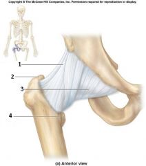
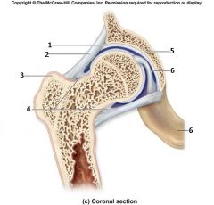
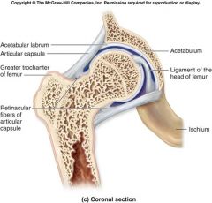
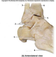
Coxal (Hip) Joint
|
a ball-and-socket joint in which a pad of fibrous cartilage covers the articular surface of the acetabulum.
|
|
|
Acetabular Labrum
|
a circular rim of fibrous cartilage increases the depth of the acetabulum.
|
|
|
Retinacular Fibers
|
The longitudinal retinacular fibers travel along the neck and carry blood vessels
|
|
|
Intracapsular Ligaments
|
|
|
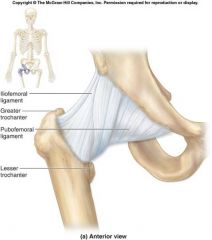
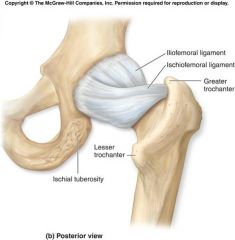
Iliofemoral Ligament
|

|
|
|
Ischiofemoral Ligament
|
|
|
|
Pubofemoral Ligament
|

|
|
|
Ligamentum Teres
|
(head of the femur) is a triangular, somewhat flattened band implanted by its apex into the antero-superior part of the fovea capitis femoris; its base is attached by two bands, one into either side of the acetabular notch, and between these bony attachments it blends with the transverse ligament
|
|
|
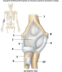
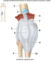
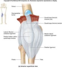
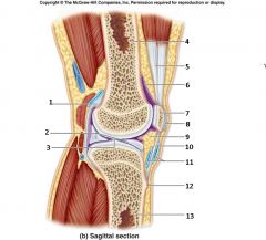
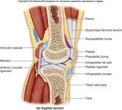
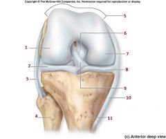
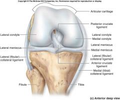
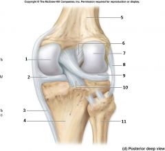
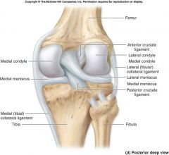
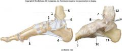
Knee Joint
|
is responsible for supporting the weight of the body along with the hip and ankle joints. Has the largest range of motion in lower limb, lacking the large muscle mass that strengthens and supports the hip, and lacks strong ligaments that support the ankle joint. It is a hinge-joint.
|
|
|
Tibiofemoral Joint
|
a joint between the femur and the tibula which helps to make up the knee joint.
|
|
|
Patellofemoral Joint
|
a joint between the patella and the patellar surface of the femur.
|
|
|
Patellar Ligament
|
provides support to the anterior surface of the knee joint where there is no continous capsule.
|
|
|
Fibular Collateral Ligament
|
reinforces the lateral surface of the knee joint. Works with the Tibial CL to tighten the extension and stabilize the joint.
|
|
|
Tibial Collateral Ligament
|
reinforces the medial surface of the knee joint. Works with the Tibial CL to tighten the extension and stabilize the joint.
|
|
|
Menisci
|
Acts as a cushion, conforms to the shape of articulating surfaces as the femur changes position, increases the surface area of the tibiofemoral joint and provide some lateral stability to the joint.
|
|
|
Menisci Medial
|

lie between the femoral and tibial surfaces.
|
|
|
Menisci Lateral
|

lie between the femoral and tibial surfaces.
|
|
|
Cruciate Ligaments
|

these ligaments limit the anterior and posterior movement of the femur and maintain the alignment of the fermoral and tibial condyles.
|
|
|
Cruciate Ligaments Anterior Cruciate Ligament (ACL)
|
refers to the anterior location on the tibia and they cross each-other as they proceed to their destinations on the femur.
|
|
|
Cruciate Ligaments Posterior Cruciate Ligament (PCL)
|
refers to the posterior location on the tibia and they cross each-other as they proceed to their destinations on the femur.
|
|
|
Costochondritis
|
is an inflammation of the cartilage that connects a rib to the breastbone (sternum). It causes sharp pain in the costosternal joint
|
|
|
TMJ Disorders
|
is an umbrella term covering acute or chronic inflammation of the temporomandibular joint, which connects the mandible to the skull. The disorder and resultant dysfunction can result in significant pain and impairment.
|
|
|
Shoulder Dislocation
|
occurs when the humerus separates from the scapula at the glenohumeral joint.
|
|
|
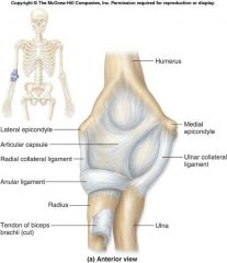
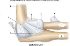
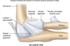
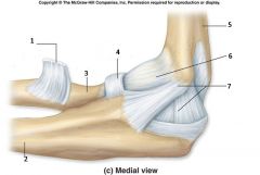
Subluxation of the Head of Radius
|
is a dislocation of the elbow joint caused by a sudden pull on the extended pronated arm, such as by an adult tugging on an uncooperative child.
|
|
|
Sprain
|
an injury to ligaments that is caused by being stretched beyond their normal capacity and possibly torn.
|
|
|
Arthritis
|
is a group of conditions involving damage to the joints of the body
|
|
|
Gouty
|
is a disease hallmarked by elevated levels of uric acid in the bloodstream. In this condition, crystals of monosodium urate (MSU) or uric acid are deposited on the articular cartilage of joints, tendons, and surrounding tissues.[1]:546 It is marked by transient painful attacks of acute arthritis initiated by crystallization of urates within and about the joints and can eventually lead to chronic gouty arthritis and the deposition of masses of urates in joints and other sites, sometimes creating tophi.
|
|
|
Osteoarthritis
|
also known as degenerative arthritis, is a group of diseases and mechanical abnormalities involving degradation of joints,[1] including articular cartilage and the subchondral bone next to it. may initiate processes leading to loss of cartilage -- a strong protein matrix that lubricates and cushions the joints
|
|
|
Rheumatoid
|
a chronic, systemic inflammatory disorder that may affect many tissues and organs, but principally attacks the joints producing an inflammatory synovitis that often progresses to destruction of the articular cartilage and ankylosis of the joints. Rheumatoid arthritis can also produce diffuse inflammation in the lungs, pericardium, pleura, and sclera, and also nodular lesions, most common in subcutaneous tissue under the skin.
|
|
|
Bursitis
|
is the inflammation of one or more bursae (small sacs) of synovial fluid in the body. When it happens movement relying upon the inflamed bursa becomes difficult and painful
|
|
|
|
|
|

|
|
|
|
|
|
|
|
|

|
|
|
|
|
|
|
|
|
|
|
|
|
|
|
|
|
|

|
|
|
|
|
|

|
|
|

|
|
|
|
|
|
|
|
|
|
|
|
|
|
|
|
|
|
|
|
|
|
|
|
|
|
|
|
|
|
|
|
|
|
|
|

|
|