![]()
![]()
![]()
Use LEFT and RIGHT arrow keys to navigate between flashcards;
Use UP and DOWN arrow keys to flip the card;
H to show hint;
A reads text to speech;
32 Cards in this Set
- Front
- Back
|
what are the layers of the scalp |
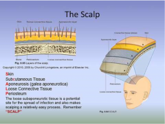
SCALP skin subCutaneous tissue aponeurosis (galea) loose connective tissue periosteum |
|
|
what are the two major muscles of the scalp |
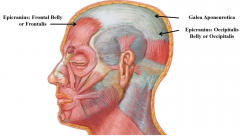
epicranius muscle (frontal and occipital belly) biventer muscle |
|
|
what are the sensory nerve branches of trigeminal nerve to the face? |
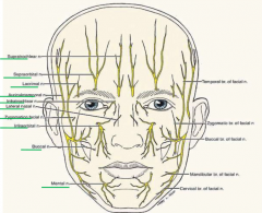
supratrochlear supraorbital lacrimal auriculotemporal infratrochlear lateral nasal zygomatic infraorbital buccal mental |
|
|
what are the sensory nerve branches of the facial nerve to the face? |
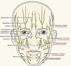
temporal
zygomatic buccal mandibular cervical |
|
|
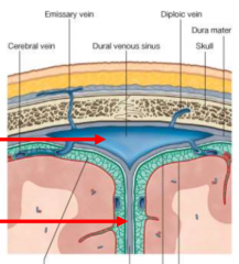
define emissary veins |
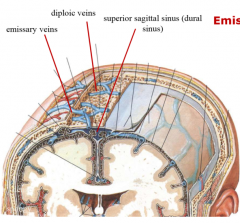
blood vessels in the scalp pass through skull and drain into dural venous sinuses |
|
|
define diploic veins |

in spongy bone |
|
|
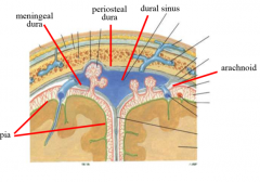
what are the 3 layers of meninges |
dura mater arachnoid mater pia mater |
|
|
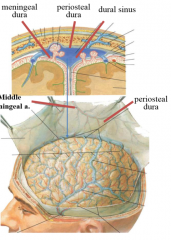
describe the dura mater |
meningeal and periosteal dura together around brain, separate around sinuses and foramen magnus (meningeal continues in spinal cord) envelops brain, spinal cord and cranial nerves arterial supply: meningeal arteries innervation: trigeminal |
|
|
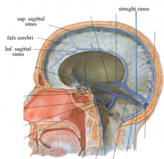
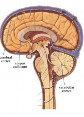
define the dura falx |
when two layers of dura mater meet and separate 2 hemispheres midline, vertically oriented partitions brain: cerebri cerebellum: cereblli |
|
|
define dural sinus |
periosteal dura adheres to the bone, meningial layer stays around brain where they split, fibrous drainage spot superior and inferior saginattal sinus simple squamous endothelium |
|
|
why do we have sinuses and not veins in the brain
|
very strong, fibrous connective tissue
intracranial pressure (cerebral spinal fluid) is higher than venous pressure, would collapse veins carries blood out of head |
|
|
define the tentorium cerebelli |
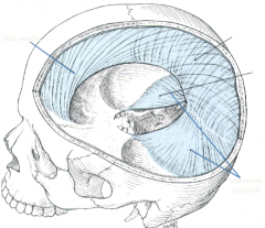
paired horizontal partitions atop cerebellar hemispheres like tents off falx cerebri at right angles straight and transverse sinus |
|
|
define the confluence of sinuses |
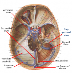
where the superior sagittal sinus comes down and meets the straight sinus goes to transverse sinuses to sigmoid sinuses out jugular foramen--> jugular vein |
|
|
define the diaphragma sellae |
circular partition roof over pituitary gland pierced by pituitary stalk |
|
|
describe the cavernous sinus |
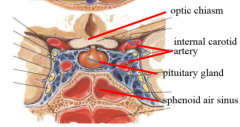
base of skull in proximity to pituitary gland contains internal carotid artery cranial nerves (III, IV, V1, V2 and VI) |
|
|
describe the course of the internal carotid artery into the head |
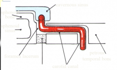
enters skull via carotid canal anteriorly and horizontally within canal enters cranium via foramen lacerum superior through cavernus sinus S shape |
|
|
what is the blood to the dura |
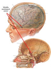
middle meningeal artery-- branch of maxillary (foramen spinosum) grooves in skull |
|
|
what is the nerve supply to the dura |
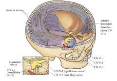
sensory from all 3 trigeminal (V1, V2, V3) middle meningeal nerve (branch of V3 via foramen spinosum) |
|
|
describe the pia mater |
delicate, transparent membrane tightly associated with the brain carries blood vessels follows folds and fissures most innermost layer |
|
|
describe the arachnoid mater |

middle layer extending from pia to dura forming subarachnoid space |
|
|
define lateral lacunae
|
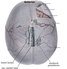
expansions of dural sinuses containing arachnoid villi |
|
|
define arachnoid granulations |
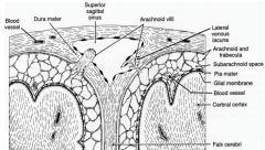
clusters of arachnoid villi absorbs cerebral spinal fluid and secretes into sinus look like cauliflower |
|
|
describe cerebrospinal fluid |
production: choroid plexus in ventricles of brain circulation: in sub arachnoid space absorption: choroid granulation into sinuses function: suspends brain, cushions it enters via median and lateral (2) apertures |
|
|
define extradural hematoma |
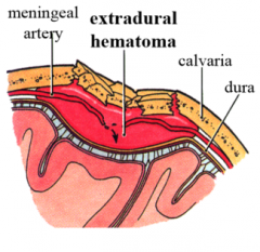
between skull and periosteal dura (skull fractures damage meningeal arteries)
localized because of adherent dura compress underlying brain tissue life threatening |
|
|
define subdural hematoma |
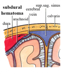
between dura and arachnoid (trauma jerks brain inside skull, tears cerebral veins as they enter sinuses) |
|
|
define subarachnoid hemorrhage |
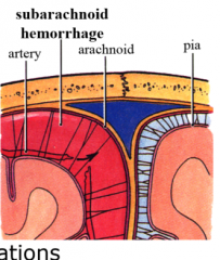
bleeding in subarachnoid space (cerebral lacerations, tearing cerebral arteries) lumbar puncture diagnostic |
|
|
describe the 3 cranial fossae |
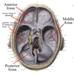
anterior: raised posterior ridge formed by lesser wing of sphenoid bone-- frontal lobes middle: raised posterior ridge, petrous crest of temporal bone-- temporal lobes and hypothalamus posterior: contains brainstem, occipital lobes and cerebellum |
|
|
describe the blood supply of the brain |
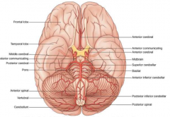
internal carotid arteries branches to middle, posterior (part of vertebral) and anterior cerebral arteries vertebral arteries from posterior cerebral and basilar arteries supply brainstem and spinal cord |
|
|
describe the circle of willis |
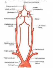
anterior and posterior communicating artery connects carotid and vertebral branches |
|
|
what are the anterior fossa cranial nerves and foraminae |
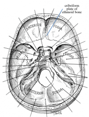
CN I-- olfactory nerve through many small holes in cribriform plate of ethmoid bone |
|
|
what are the middle fossa cranial nerves and foraminae |
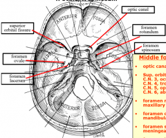
CN II-- optic nerve through optic canal CN III oculomotor/CN IV trochlear/CN V1 trigeminal (ophthalmic)/CN VI abducens through superior orbital fissure CN V2 trigeminal (maxillary) through foramen rotundum CN V3 trigeminal (mandibular) through foramen ovale middle meningeal artery and nerve through foramen spinosum greater petrosal nerve (part of CN VII) through foramen lacerum |
|
|
what are the posteriro fossa cranial nerves and foraminae |
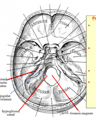
CN VII facial/CN VIII vestibulocochlear through internal acoustic meatus CN IX glossopharyngeal/CN X vagus/ CN XI accessory (plus internal jugular vein) through jugular foramen CN XII hypoglossal through hypoglossal canal medulla oblongata (with vertebral arteries and spinal accessory nerve) through foramen magnum |