Reading...
![]()
Play button
![]()
Play button
![]()
Use LEFT and RIGHT arrow keys to navigate between flashcards;
Use UP and DOWN arrow keys to flip the card;
H to show hint;
A reads text to speech;
67 Cards in this Set
- Front
- Back
|
Four routes of infection of the CNS
|
1. Bloodstream- most common portal of entry
2. Direct Implantation- usually traumatic 3. Local Extension- usually from sinuses (bad teeth) 4. Peripheral Nervous System- conduit for some viruses, especially rabies and Herpes Simplex |
|
|
3 main infections of the CNS
|
-Meningitis- Infection of meninges and CSF
-Encephalitis- Infections of brain parenchyma -Meningoencephalitis- Infection of meninges and brain parenchyma |
|
|
What are the central hallmarks of acute pyogenic meningitis?
|
• Increased pressure
• Increased polymorphonuclear leukocytes • Increased protein • Decreased glucose It is a bacterial meningitis that is result of hematogenous spread |
|
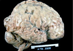
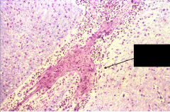
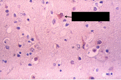
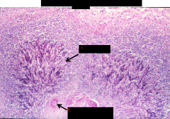
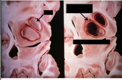
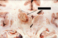
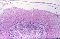
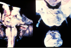
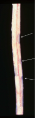
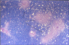
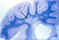
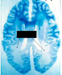
What infection is this a picture of?
|
Acute Pyogenic Meningitis
note the pus in the subarachnoid space that sometimes can penetrate into the Virchow-robin spaces of the brain the blood vessels are surrounded and this --> vasculitis |
|
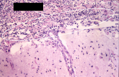
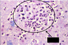
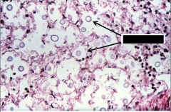
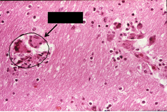
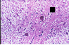
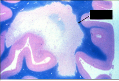
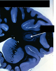
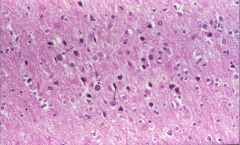
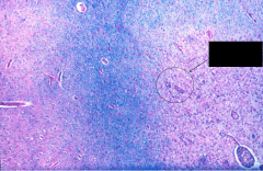
What is depicted here?
What is indicated by the white circle? |
Acute Pyogenic Meningitis
White circle: neutrophilic infiltration Lots and lots of leukocytes Give rise to vasculitis and infarction Doesn't happen if treated properly |
|
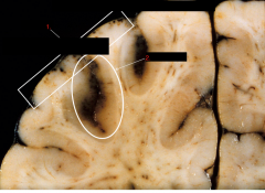
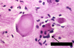
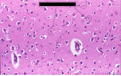
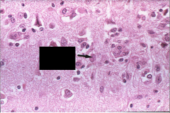
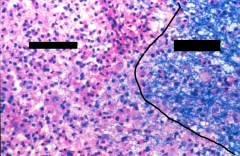
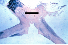
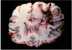
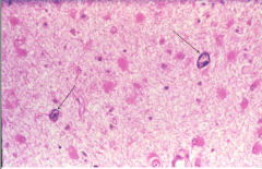
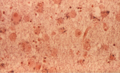
What disease does this depict?
What are 1. and 2.? |
Acute Pyogenic Meningitis
1. Vasculitis 2. Hemorrhage |
|
|
What are the most common causes of Acute Pyogenic Meningitis?
|
- Neonate - Escherichia coli
• Infants and Children - Hamemophilus influenza • Adolescents and Young Adults - Neisseria meningitidis • Adults and Post-Traumatic - Pneumococcus |
|
|
What are the diagnostic hallmarks of Acute Lymphocytic Meningitis?
|
- Lymphocytic pleocytosis
• Moderate protein elevation (< than bacterial) • Glucose nearly normal - Viral load increased Associated with viral infections of the brain, mononuclear infiltrate in the CSF |
|
|
Pathologic hallmarks of Acute Lymphocytic Meningitis?
|
• perivascular and parenchymal mononuclear cell infiltrates (lymphocytes, plasma cells and macrophages)
• glial nodules (microglia that begin to digest the dead neurons) • neuronophagia • inclusion bodies (viral factories within cells that have been infected) |
|
What disease does this depict? What causes this disease?
|
- Viral Meningoencephalitis
- Caused by Eastern Equine virus - in a boy scout - camping trip on block island - brain swollen, not a lot of exudate, vessels congested; others were exposed, but he was the only one who died. Immune systems can fight it -vasculature is dilated |
|
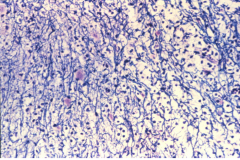
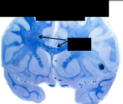
What disease is depicted here? What is the arrow pointing to?
|
Viral Meningoencephalitis
Arrow points to: Perivascular lymphocytic Infiltrate |
|
What does this image depict? What disease is present here?
|
Depicts neuronophagia, in which glial cells eat infected/dying neurons
Viral Meningoencephalitis |
|
What does this depict What disease would you see this in?
|
From Viral Meningoencephalitis - this is a microglial nodule, evidence of prior neuronophagia (glia digesting infected/dying neurons)
|
|
What is depicted at the arrow? What disease is this evidence of?
|
Eosinophilic Nuclear Inclusion, aggregates of viruses using host DNA to reproduce itself
Evidence of Viral Meningoencephalitis |
|
What is depicted here? What disease is this?
|
This is a cell stained positively for Herpes Virus
Cause of viral meningoencephalitis |
|
|
Where does chronic meningitis usually occur? What does it cause?
|
• Base of brain extending into the lateral sulci
• Obliterative endarteritis of arteries in subarachnoid space--> hyperplasia that leads to arterial occlusion and infarction |
|
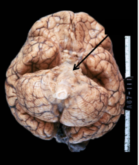
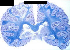
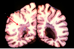
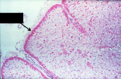
What disease is depicted here? What is the arrow pointing to?
|
- Chronic meningitis
- Black arrow = basilar exudate - Unlike acute meningitis, where you have sugar coating in convexities, you have this at the base of the brain - far less dramatic in most instances The disease will produce obliterative endarteritis in subarachnoid space |
|
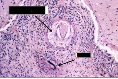
What disease is depicted here? What is indicated by arrows 1. and 2.?
|
- this is found in chronic meningitis
1) Obliterative Endarteritis with Intimal Hyperplasia 2) Giant Cell, produced from granulomous inflammation This is an inflammatory infiltrate that is granulomatous; lymphocytes + plasma cells + foreign body giant cells These can secrete things that make endothelial cells proliferate and block BV --> infarction |
|
What disease is depicted here?
|
Fungal meningitis - a very severe case
Cryptococcal meningitis Common cause of chronic meningitis |
|
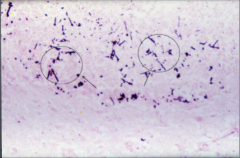
What disease is depicted at the arrows?
|
Cryptococcus - cryptoccocal meningitis (fungus)
Common cause of chronic meningitis |
|
What disease is depicted here?
|
This is Candida Albicans - chronic meningitis found in immunocompromised individuals
|
|
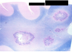
What disease is this? What is indicated in the circles?
|
Candida Albicans (chronic meningitis)
Black circles = pseudohyphae |
|
What disease is this?
|
Aspergillus (chronic meningitis)
Found in immunocompromised people SEe vascocentric necrosis and hemorrhage |
|
What disease is this? What is depicted at 1. and 2.?
|
Aspergillus (chronic meningitis)
1. True Hyphae 2. Necrotic Blood Vessels This disease usually only infects those that are immunocompromised |
|
|
What causes brain abscesses? What are the pathological hallmarks of brain abscesses?
|
• direct implantation, local extension or hematogenous spread
• associated with acute bacterial endocarditis, cyanotic congenial heart disease and chronic plumonary sepsis • anaerobic streptococci, Bacteroides fragilis, aerobic streptococci and staphylococci most common pathogens • focus of necrotizing cerebritis • fibrous capsule produced by fibroblasts derived from blood vessels • zone of gliosis outside fibrous capsule A local infection where the body walls it off, creates fibrous capsule to wall it off |
|
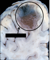
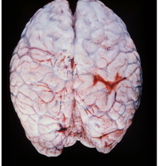
What disease process is depicted here?
|
Brain Abscess
|
|
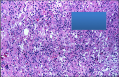
What disease process is present here? What cells are present?
|
Brain abscess
Absess filled with polymorphonuclear leukocytes, macrophages Pool formation of reticulin, congeals into fibrous capsule |
|
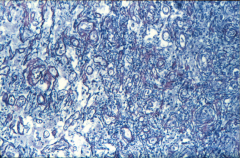
What is depicted here? What disease process is this indicative of?
|
Reticulin in blood vessels; this proliferates to wall off pus from a BRAIN ABSCESS from the rest of the brain
|
|
What is indicated by the arrow on the left? What about the arrow in the right?
|
L: Pus (this is a brain abscess)
R: Fibrous Capsule left behind after pus removal This could be a location of seizure focus |
|
|
What are 4 syndromes caused directly by HIV?
|
1. acute meningitis or encephalitis
2. subacute encephalitis 3. vacuolar myelopathy 4. peripheral neuropathy (but we rarely see these because of the treatment of HIV now) No inclusion bodies |
|
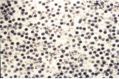
What disease is this indicative of? What is circled in black?
|
These are multinucleated giant cells of monocytic origin which are hallmarks of HIV meningitis
|
|
What disease is this indicative of? What is circled in black?
|
These microglial "stab" cells are found in HIV and syphilis-related meningitis
Stab cells are slender knife-like microglial cells |
|
What is depicted here?
|
Viral particles within an infected neuron
|
|
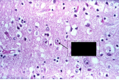
What is depicted at the arrow?
|
Toxoplasmosis - found in patients who are immunocompromised; this is a secondary infection in the brain
Has mulberry appearance in toxo cyst |
|
What is depicted at the arrow?
|
This is Toxoplasmosis
At the arrow, you see "Mulberry" appearance of a toxo cyst |
|
What is depicted at the arrow?
|
A toxo cyst; as seen in Toxoplasmosis
|
|
|
List 5 prion diseases.
|
• Creutzfeldt-Jakob Disease (sporadic and hereditary)
•Kuru •New Variant CJD (Mad Cow Disease) •Fatal Familial Insomnia (hereditary) •Gerstmann - Straussler Disease (hereditary) |
|
|
Creutzfeld-Jakob disease: symptoms, pathology, and etiology
|
• Rapidly progressive dementia with myoclonus and periodic EEG pattern
• Spongiform encephalopathy with “kuru” plaques in 5-10% • Etiology thought to be accumulation of abnormal “prion” protein, cause other proteins to misfold Both sporadic and hereditary |
|
What disease is this?
|
Cruetzfeldt-Jakob Disease
• Rapidly progressive dementia with myoclonus and periodic EEG pattern • Spongiform encephalopathy with “kuru” plaques in 5-10% • Etiology thought to be accumulation of abnormal “prion” protein, cause other proteins to misfold Both sporadic and hereditary |
|
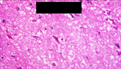
What disease is depicted here? What are the white bubbles indicative of?
|
CRUETZFELDT- JAKOB DISEASE
Prion protein accumulates, forming white bubbles |
|
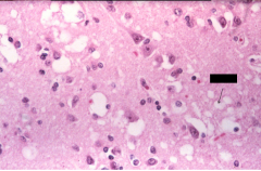
What disease is depicted here? What is at the arrow?
|
CRUETZFELDT- JAKOB DISEASE
Arrow: Prion protein accumulates, forming white bubbles |
|
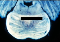
What is depicted here?
|
Spongiform Transformation in molecular layer of the cerebellum
Can occur because of prion diseases |
|
What disease is depicted here?
|
Multiple Sclerosis: Demyelinating Disease
You can see the sharply demarkated area of demyelination (red arrow) Demyelination usually perivenous- formation of plaques |
|
What is depicted here?
|
MS plaques
Demyelination usually perivenous Seen often in the cerebrum, optic, brainstem, and spinal cord |
|
What disease is this? What is depicted at the arrow?
|
Multiple Sclerosis
Arrow: Area of demyelination Sharp line where demyelination occurs |
|
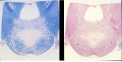
What disease is this? Which side is myelinated and which side is demyelinated? Which is normal
|
MS
L: abnormal, demyelinated R: normal myelin |
|
What is notable about this slide? What disease is it?
|
MS - notable because axons are preserved, but myelin is missing
|
|
|
What are the morphological features of MS?
|
Topography
1. Cerebrum, optic, brainstem, spinal cord 2. Predominately white matter with grey matter affected 3. Asymmetric, discontinuous and unsystemized 4. Periventricular and subcortical 5. Sharp demarcation of grossly intact tissue Histopathology 1. Perivascular (venous) 2. Oligodendrocytes diminished 3. Perivascular and meningeal mononuclear infiltrate 4. Myelin loss 5. Axons spared 6. Macrophage formation with sudanophilic breakdown 7. Concentric spread |
|
What disease is this?
|
Variant of MS: Devic's disease
Demyelination limited to optic chiasm and spinal cord Defect in aquporin 4 |
|
What disease is this? What is indicated at the arrows?
|
MS in the optic chiasm
The arrows show demyelination |
|
What disease is this? What is indicated at the arrow?
|
MS Variant: Balo's Concentric Sclerosis
Arrow: Concentric lesion in cerebral white matter Tiger striped lesion is distinct |
|
What is depicted here? What is indicated at the arrows?
|
Post-Infectious Encephalomyelitis or ADEM: Acute Disseminated Encephalomyelitis
Arrow: multi-focal perivenular demyelination This is LIKE MS, but gets better; found in the context of a previous infection ADEM is an autoimmune disease usually triggered by viral infection, but can also occur due to vaccine Note: caused because health Nazis make us get flu shots. |
|
What disease is depicted here? What is at the arrow?
|
ADEM: Acute Disseminated Encephalomyelitis
Arrows: Perivascular Demyelination |
|
What is depicted here?
|
Acute hemorrhagic leukoencephalitis
Can occur from demyelination (like from ADEM) when it becomes hemorrhagic (fatal) |
|
|
Antecedent events for Guillain-Barre Syndrome
|
1.VIRAL INFECTIONS Measles
Mumps Rubella Influenza A Influenza B Varicella-zoster Cytomegalo virus Epstein-Barr virus Infectious mononucleosis Vaccinia Variola Infectious hepatitis Coxsackie ECHO 2. Other infections Mycoplasma pneumoniae Salmonella typhi Camphylobacter jejuni Listerosis Brucellosis Tularemia Ornithosis 3. VACCINES Rabies Swine Flu 4. Surgery 5. Fever therapy 6. MALIGNANCY Hodgkin’s disease Carcinoma Lymphoma 7. PREGNANCY USUALLY PRESENTS AFTER GASTROINTESTINAL ILLNESS |
|
What is depicted here? What causes this?
|
Central Pontine Myelinolysis
Death of oligodendrocytes and corresponding loss of myelin in the pons cause: rapid correction of hyponatremia Can --> quadriplegia |
|
What is depicted here?
|
Central pontine myelinolysis
Death of oligodendrocytes and corresponding loss of myelin in the pons cause: rapid correction of hyponatremia Can --> quadriplegia Axons are preserved and inflammation is absent |
|
What is this a picture of?
|
Progressive Multifocal Leukoencephalopathy (PML)
Caused by papovaviridae- usually in immunocompromised patients |
|
What is depicted here?
|
Oligodendroglial nuclear inclusions
Characteristic of progressive mulitfocal leukoencephalopathy |
|
What is depicted here?
|
"Bizarre astrocytes"
Characteristic of progressive mulitfocal leukoencephalopathy |
|
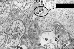
What is depicted here?
|
JC virus on EM
We all have this virus, all kept in check Usually limited to immunocompromised patients |
|
What is depicted here?
|
Adrenoleukodystrophy
Arrows point to areas of dysmyelination Failure of normal myelin to form This leads to macrophages to digest the abnormal myelin |
|
What is depicted here? What is at the arrow?
|
Adrenoleukodystrophy
Failure of normal myelin to form Macrophages digest the abnormal myelin |
|
What is depicted here?
|
Metrachromatic Leukodystrophy
|
|
What is depicted here? What is at the arrow?
|
Alexander's Disease
Arrow: Rosenthal fibers |
|
What disease is depicted here?
|
Tay Sach's disease
A storage disease in which there is a deficiency in hexosaminadase A Leads to the death of neurons, kills children from a young age |
|
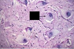
What disease is this? What is at the arrow?
|
- Tay Sach's
- Swelling in the neuronal cytoplasm From the buildup of lysosomal products that cannot be broken down since there is a deficiency of hexosaminadase A Leads to death of neurons and early childhood death |