![]()
![]()
![]()
Use LEFT and RIGHT arrow keys to navigate between flashcards;
Use UP and DOWN arrow keys to flip the card;
H to show hint;
A reads text to speech;
55 Cards in this Set
- Front
- Back
|
germ layer that forms epidermis of the skin, epithelium of the oral and nasal cavities, and nervous system and sense organs.
|
Ectoderm
|
|
|
germ layer that forms mucosal epithelium and glands of respiratory and digestive systems.
|
Endoderm
|
|
|
germ layer that forms muscle and connective tissue, including bone, and components of the
circulatory, urinary and genital systems. |
Mesoderm
|
|
|
primitive gut tube is derived from _________
(be specific; what structures involved and how is formed) |

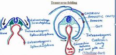
portion of endoderm-lined yolk sac cavity (surrounds/engulfs y.sac), which is incorporated into the body of the embryo during tranverse folding of the embryo (during fourth week in human).
note: primitive gut tube that forms is longitudal tube; lateral folds, covered with somatopleure (somatic lateral mesoderm + ectoderm), migrate along transverse plane to pinch off yolk sac |
|
|
primary tissue of the digestive tubes (like epithelium and glands) are derived from?
whereas most CT and muscular tissue of dig.system is derived from? |
endoderm
splanchnic mesoderm |
|
|
Foregut forms what structures?
Suppled by what artery? |
Supplied by Celiac artery (branches of celiac trunk)
Forms pharynx, lower respiratory system, esophagus, stomach, proximal half of duodenum, liver and pancreas, biliary apparatus |
|
|
Midgut forms what structures?
Suppled by what artery? |
Supplied by superior mesenteric artery
Small intestine, distal half of duodenum, cecum and vermiform appendix, ascending colon, most of the transverse colon |
|
|
Hindgut forms what structures?
Suppled by what artery? |
Supplied by Inferior mesenteric artery
Left part of transverse colon, descending colon, sigmoid colon, rectum, superior part of anal canal, epithelium of urinary bladder, most of the urethra |
|
|
liver and pancreas are derived from _____
|
endoderm
|
|
|
muscular walls of the digestive tract (lamina propria, muscularis mucosae, submucosa, muscularis externa, adventitia and/or serosa) are derived from _______?
|
splanchnic mesoderm
|
|
|
(anal pit) is the primordial anus is known as the __________?
primordial mouth is known as? ------------------------ Challenge Question: germ layer present? how do these form? |

proctodeum
stomodeum In both of these areas ectoderm is in direct contact w/endoderm w/o intervening mesoderm, eventually bucco-pharyngeal and cloacal membranes degenerate forming a continuous digestive tube from the mouth to the anus |
|
|
At birth, how do reticulum and rumen combined compare in size to abomasum?
At 8 wks, reticulum and rumen compare to abomasum? At 12 weeks, how do reticulum and rumen (combined) of calf compare in size to abomasum? Describe growth of omasum? |
half size (collapsed, not functional on milk diet)
At 8 wks, reticulum/rumen = abomasum By 12 weeks, abomasum twice size very slow, in adult same size as abomasum |
|
|
The _________ is largest of ruminant chambers, making up at least 75% of stomach capacity.
Smallest in large ruminant? Smallest in small ruminant? |
Rumen
Reticulum (5% in LR) Omasum (4% in SR, but 8% in LR) |
|

During midgut, as primary intestinal loop lengthens, the _____loop grows faster than the ______loop causing twisting around _____.
|

the CRANIAL loop grows faster than the caudal loop causing twisting around Cranial Mesenteric Artery
|
|
|
urorectal septum seperates
(looking for specific term used to describe ventral structure) |

ventral UROGENITAL SINUS from dorsal rectum
continues caudally to separate anal canal and external genitalia |
|
|
terminal end of dorsal rectal septum is called
|

perineal region
|
|
|
Atresia ani?
What animals common in? |

Abnormalities
(imperforate anus) failure of anal membrane to break down in calves and pig. |
|
|
Counter-rotation of gut
What side does duodenum end up on? What side does Desc. colon end up on? |
Abnormalities
(Situs invertus) when the body organs develop opposite to their normal position e.g. descending duodenum on the left and descending colon on the right. |
|
|
urine in the rectum?
urine out the bellybutton? |
Urorectal fistula
note: a fistula is abnormal connection or passageway between two epithelium-lined organs or vessels that normally do not connect; generally a disease condition, but a fistula may be surgically created for therapeutic reasons. patent urachus- connection of the allantois to the urinary bladder will become the urachus. |
|
|
midgut intestinal loop failed to retract, produces a congenital hernia
(intestine sticking out umbilicus) |

Omphalocele
|
|
|
Hepatic diverticulum (name 2 structures budding off stomach there)
|
Pars hepatica and pars cystica (forms GB)
|
|
|
Left lobe of pancreas develops from?
Right lobe – Pancreatic duct develops from? |

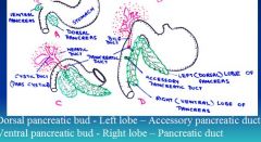
Dorsal pancreatic bud (accessory pan. duct - absent in sm.ruminant)
Ventral pancreatic bud (pancreatic duct - absent in ox and pig) |
|
|
Which animal has large pancreatic duct (orig.ventral)?
Absent in? |
Horse/Cat/Sm.Ruminant
Ox/Pig |
|
|
Which animal has large accesory duct (orig.dorsal)?
Absent in? |
Dog
Sheep/Goat |
|
|
_______ develops from mesoderm of the dorsal mesogastrium (from primitive dorsal mesentery)
|

spleen; its above stomach
|
|
|
epiploic foramen and omental bursa develops b/c of ?
|

rotation to foregut or stomach along longitudinal axis
|
|
|
vitelline duct connects what?
|
vitelline duct or yolk stalk connects MIDGUT and yolk sac
|
|
|
how does greater curvature form?
|

dorsal wall of developing stomach grows faster than ventral wall
|
|
|
At cranial most end of foregut, ______dermal depression known as ______ develops.
|
Cranial most end of foregut, EXTERNAL ECTODERMAL depression known as the STOMODEUM develops.
|
|
|
somatopleure?
splanchnoplure? |

somatic mesoderm + ectoderm
splanchnic mesoderm + ectoderm |
|
|
describe the 2 rotations of dev stomach?
|

first rotation along cranio-caudal axis, moves greater curvature to left
stomach and duodenum rotate along ventrodorsal (A-P) axis pulling end of stomach upwards |
|
|
Proportion of each stomach compartment in (adult) large ruminant?
|
Rumen: 80%
Reticulum: 5% Omasum: 8% Abomasum: 7% Note: abomasum is similar in anatomy and function as the monogastric stomach |
|
|
Proportion of each stomach compartment in small ruminant (adult) ?
|
Rumen: 75%
Reticulum: 8% Omasum: 4% Abomasum: 13% |
|
|
How does endoderm become continuous with ectoderm at stomodeum and proctodeum?
|
bucco-pharyngeal and cloacal membranes rupture
forms the continuity of digestive tube from mouth to anus |
|

identify this structure
what does it form? |

cranial limb
it forms small intestine |
|
|
Why does intestinal loop enter extraembryonic coelon?
What does caudal limb of primary intestinal loop form? |
Because of insufficient space in abdominal cavity
Lower part of ileum, cecum, ascending and part of transverse colon |
|
|
blood supply to primary intestinal loop?
|
superior mesenteric artery
|
|
|
which intestine loop grows more quickly?
|

Cranial loop grows more rapidly than caudal, causing rotation
(around cranial mesenteric artery) |
|
|
the pyloric part of developing cow stomach forms the ______?
|
abomasum
|
|
|
In _______ asc.loop of colon is short straight, followed by transverse and desc.colon.
In _______asc.loop of colon is elongated, which grows into proximal and distal and SPIRAL loops. |

dog and cat (asc.loop of colon is short straight)
ruminant = spiral loops of asc.colon |
|
|
In pig asc.colon develops into _____ coils
|

In pig asc.colon develops into CENTRIPETAL and CENTRIFUGAL coils
|
|
|
urorectal septum derived from?
|
endoderm and thickening of surrounding mesenchyme
|
|
|
liver develops as ventral outgrowth from _______ at terminal part of _______.
|
liver develops as ventral outgrowth from GUT ENDODERM at terminal part of FOREGUT
|
|
|
Hepatic bud or diverticulum which consists of rapidly prolif. cells develops into what two parts? (which one is absent in horses)
Which develop into? |
Pars HEPATICA: dev. into liver parenchyma (liver cords) and hepatic ducts
note: parenchyma is functional part of an organ Pars CYSTICA: will form gall bladder and cystic duct (this one is absent in horses) |
|
|
During further development, epith.liver cords mix w/vitelline veins to form ______?
Liver cords differentiate into ____? |
liver cords + vitelline veins = hepatic sinusoids
Liver cords (which are branching and proliferating) differentiate into hepatocytes....this establishes the characteristic hepatic lobules. |
|
|
kupffer cells are derived from?
|
kupffer cells and CT of liver are derived from MESODERM of SEPTUM TRANSVERSUM
note: kupffer cells are specialized macrophages located in liver lining the walls of sinusoids |
|
|
accessory pancreatic duct enters duodenum at site of ______ diverticulum.
|

accessory pancreatic duct enters duodenum at site of DORSAL pancreatic diverticulum.
note: picture from notes might be wrong b/c shows acc.duct coming from ventral diverticulum |
|
|
accessory pancreatic duct is prominent in ?
small in? absent in? |
accessory pancreatic duct is prominent in DOG, OX, PIG
but small in horse & cat absent in small ruminant |
|
|
Glanzmann's dz
|
AR
GpIIb/IIIa receptor deficiency for fibrinogen Defective platelet aggregation increased bleeding time |
|
|
laryngotracheal diverticulum arises from _________?
|

endoderm on the ventral wall of the foregut
|
|
|
Tracheoesophageal folds develop on either side and join to form ________ that separates it from the rest of the foregut.
|

tracheoesophageal septum
This divides the foregut into the laryngotracheal tube (ventral) and the esophagus (dorsal). |
|
|
The caudal end of the laryngotracheal diverticulum enlarges to form the lung bud, which is surrounded by _______.
|
splanchnic mesenchyme
|
|
|
The respiratory diverticulum (lung bud) appears as an outgrowth from the ______?
So epithelium lining larynx, trachea, bronchi and lungs is entirely ____? cartilaginous and muscular component are derived from ____ surrounding ______. |
ventral wall of the foregut.
endodermal splanchnic mesoderm, mesoderm |
|
|
formation of pleuropericardial septum (mediastinum, etc)
|
at first heart is beneath lung buds
seperation betw. pleural cavity and heart related to growth of 2 common cardinal veins on either side of lung buds, mesench.tissue around c.c.v. (called pleuropericardinal fold) starts to move toward eachother, and eventually fuse this seperates growing lungs and forms ppc septum. beneath allow heart to rise betw. them As lung grow, this horiz.septum expands |
|
|
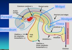
primitive gut tube is divided into three sections.
|
Foregut
midgut hindgut |

